Etiketter

Visar inlägg med etikett MMP-9. Visa alla inlägg

fredag 27 oktober 2017

Lymen neuroborrelioosi ja liquorin MMP-kirjo

https://www.ncbi.nlm.nih.gov/pubmed/?term=TIMP-1+in+Lyme+neuroborreliosis

J Neurol Neurosurg Psychiatry. 2000 Mar;68(3):368-71.

Upregulation of matrix metalloproteinase-9 in the cerebrospinal fluid of patients with acute Lyme neuroborreliosis.Kirchner A1, Koedel U, Fingerle V, Paul R, Wilske B, Pfister HW

Abstract

It was investigated
 (1) whether metalloproteinase-9 (MMP-9), MMP-3, and tissue inhibitor of matrix metalloproteinase-1 (TIMP-1, the natural tissue inhibitor of MMP-9) are increased in the CSF of patients with Lyme neuroborreliosis and
(2) whether macrophages can express MMP-9 when stimulated with Borrelia burgdorferi. Zymography showed MMP-9 activity in 26 of 31 (84%) CSF samples from patients with acute stage 2 Lyme neuroborreliosis, but not in 20 controls with non-inflammatory neurological disorders.

 Activity of MMP-2 was detected in all CSF samples in both patients with neuroborreliosis and controls, suggesting a constitutive release of MMP-2.

Using enzyme linked immunosorbent assay (ELISA) MMP-3 (which can activate MMP-9) was detected in low concentrations in the CSF of 13 of 29 patients with neuroborreliosis, but not in controls.

TIMP-1 was increased twofold in CSF samples from patients with neuroborreliosis in comparison with the controls.

MMP-9 activity was induced in vitro in a mouse macrophage cell line (RAW 264.7) when stimulated with two different genospecies of B burgdorferi (B garinii, B afzelii ). This MMP-9 activity was reduced in a dose dependent manner when macrophages stimulated with B burgdorferi were coincubated with NF-kappaB SN50, a cell permeable peptide which inhibits the translocation of NF-kappaB into the nucleus of stimulated cells.

 The data show that (1) MMP-9 activity is present in the CSF of patients with neuroborreliosis,
 (2) macrophages stimulated with B burgdorferi are a possible source of MMP-9 increase, and
(3) activation of NF-kappaB may play a part in the upregulation of MMP-9 by B burgdorferi.
PMID:
10675223
PMCID:
PMC1736835
[Indexed for MEDLINE]
Free PMC Article

fredag 19 september 2014

MMP, TIMP ja MT-MMP järjestelmä syövän invaasiossa- purkamassa basaalikalvoa . Kirjon erilaisuus primäärisyövässä ja sen ihometastaasissa

Alaloleva tilanne  kuvaa  matrixmetalloproteinaasijärjestelmän molekyylejä MMP, TIMP ja  myös MT-MMP  työssään hajoittamassa ECM , extrasellulaarimatrixia ja täten  autamassa syöpäsolua  läpi  basaalikalvosta strooman puolelle.( esim  juuri  EMT ilmiössä  epiteeli-mesenkyymi- transitiossa. 

http://www.ncbi.nlm.nih.gov/pubmed/25062266
Am J Dermatopathol. 2014 Jul 24. [Epub ahead of print]
Comparative Expression of Matrix Metalloproteinases in Internal Malignancies and Paired Cutaneous Metastatic Lesions.
Tiivistelmä Abstract
  • Tausta:
On ajateltu, että matrixmetalloproteinaasi (MMP) ja metalloproteinaasin kudosestäjä (TIMP) sekä membraanityyppinen matrixmetalloproteinaasi 1 ( MT1-MMP) osallistuvat peruskalvonbasaalimembraanin  tuhoamiseen ja syöpäsolujen invasoitumiseen strooman puolelle.

BACKGROUND:
Matrix metalloproteinase (MMP), tissue inhibitor of metalloproteinase (TIMP), and membrane-type 1 matrix metalloproteinase (MT1-MMP) are thought to be involved in the destruction of basement membrane and stromal invasion by cancer cells.

  • Tämän työn tarkoitus:
Työssä on koetettu identifioida, tunnistaa ja vertailla MMP ja TIMP- ilmentymisiä erilaisissa  sisäelinten pahanlaatuisissa taudeissa ja ihon metastaattisissa vaurioissa.

OBJECTIVE::
The aim of this study was to identify and compare MMP and TIMP expression in internal malignancies and paired cutaneous metastatic lesions.

  • Aineisto ja menetelmät:
Vertailtiin  entsyymit  MMP-2, MMP-9, MT1-MMP ja TIMP-2 sisäelinten  maligniteeteissa ja  niitten vastaavissa  ihometastaasi leesioissa käyttämällä immunohisokemiallista värjäystä. 

MATERIALS AND METHODS::
We compared the expression of MMP-2, MMP-9, MT1-MMP, and TIMP-2 in the internal malignancy and paired cutaneous metastatic lesion using immunohistochemical stains.

  •  Tulokset:
 Pahanlaatuisissa iholeesioissa ilmeni merkitsevästi enemmän MMP-2, MMP-9 ja MT1-MMP entsyymejä ja merkitsevästi vähemmän  inhibiittoria  TIMP-2 kuin mitä havaittiin vastaavassa  elinmaligniteeteissa.
Ca Mammae, Rintasyövässä ihon metastaattisissa leesioissa ilmeni  merkitsevästi enemmän MMP-9 ja merkitsevästi vähemmän TIMP-2 kuin mitä havaittiin primäärissä rintasyöpäleesiossa.
Ca pulmonalis, Keuhkosyövässä ihon metastaattisissa leesioissa oli merkitsevästi enemmän  MMP-2 ja MT1-MMP kuin primäärileesiossa.
Ca ventriculi,Mahasyövässä vastaavat ihomaligniteetit  ilmensivät merkitsevästi vähemmän TIMP-2 estäjäentsyymiä mitä  taas primäärileesio.

RESULTS:
The cutaneous metastatic lesions expressed significantly more MMP-2, MMP-9, and MT1-MMP and significantly less TIMP-2 than did the paired internal malignancies. In breast cancer, cutaneous metastatic lesions expressed significantly more MMP-9 and significantly less TIMP-2 than did the primary breast cancer lesion. In lung cancer, the cutaneous metastatic lesion expressed significantly more MMP-2 and MT1-MMP than did the primary lesion. In stomach cancer, the cutaneous metastatic lesion expressed significantly less TIMP-2 than did the primary lesion.

  • Johtopäätökset:
 Tutkijaryhmän johtopäätöksena oli, että metastaattisissa ihovaurioissa oli erilainen  MMP- ja TIMP_2 ilmentymäkirjo verrattuna  niitä vastaaviin  (sisä)elinmaligniteettiin.Lisäksi MMP ja TIMP-2  kirjot ovat  eri primäärisyöpätyypeissäkin erilaiset.

CONCLUSIONS::
Our study demonstrates that cutaneous metastatic lesions have different MMPs and TIMP-2 expression patterns compared with their paired internal malignancies. Also, MMPs and TIMP-2 expression differs according to the type of primary cancer.
PMID:
25062266
[PubMed - as supplied by publisher]

Päivitys 14.11. 2018
Kts. MT1-MMP osuus MUC1 signaloinnissa . Muc 1 säätyy ylös  monesa syövässä, mikä edistää syövän kasvua, ja progredioitumsita.
MUC1 tietoa lisää:

Cell Death Dis. 2014 Oct; 5(10): e1438.
MUC1 irtoaminen ”lehteily” tai ”hilseily” epiteelisolusta tyviosansa kompleksista ei tapahdu ilman säätelyä ja siinä säätelevät ” sheddase” entsyymit ovat MT-MMP ja TACE.

fredag 18 april 2014

Michael Jonssonin väitöskirjasta edelleen : Aivojen valkean aineen muutoksista kognition osoittaessa heikentymää

Aivojen valkea aines muuttuu henkilöillä, joilla on kognitiivista heikentymää. kliinisiä ja patologisia näkökohtia. Micghael Jonsson on ottanut biomarköörien joukkoon minua erityisesti kiinnostavan  molekyylin, sulfatidin, sillä kaikki aivosulfatidit vaativat syntetisoitumiseksiin  K-vitamiinin ja B6 vitamiinin balansoitua yhteistyötä.  Sulfatidien vähyys näkyy olevan  axonaalisessa  degeneraatiossa ilmeistä( myeliinin pirstoutumisessa). Siis tässä voi ainakin  toivoa, että vihreä, kasvispitoinen  K-vitamiinipitoinen ravinto ja K-vitamiinipitoiset kasvisöljyt eivät puuttuisi  ikääntyvien jokapäiväisestä ravinnosta.K-vitamiinilla valkeassa aineesa onmyös anti-infektiöösi vaikutus  kertoo eräs tutkimus toisessa yhteydessä.

Avainsanoja Nyckelord : 

Yhteenveto:Aivojen valkean aineen muutokset (WMC) , jotka on havaittu  tietokonetomografialla tai magneettiresonanssikuvauksella, edustavat  iäkkäillä ihmisillä  tavallista tilaa.  Kuitenkin prognostiset, symptomatologiset ja biokemialliset osatekijät  tässä valkean aineen muuntumisessa on vain ositatin  tiedossa.  Tähän kohtaan pureutuu M Jonssonin väitöskirja. 

Sammanfattning :

. The aim of the present study was to evaluate WMC in relation to clinical manifestations in patients with mild cognitive impairment (MCI) and dementia and, by means of cerebrospinal fluid (CSF) analyses, to study different structural biomarkers possibly reflecting the pathophysiological process of WMC in non-disabled patients.

 In study I, significant associations were found between WMC and age, sex, hypertension, ischemic heart disease and TIA/minor stroke. Furthermore there were significant associations between WMC and apathy, mental slowness, disinhibition, gait disturbance and focal neurological symptoms, but not with depressed mood.

 In study II, CSF was analyzed for biomarkers known to be related to Alzheimer’s disease [AD; the 1-40 and 1-42 fragments of amyloid-β, α- and β-cleaved soluble amyloid precursor proteins (sAPPα, sAPPβ), total tau (T-tau), hyperphosphorylated tau (P-tau)] and vascular dementia [VaD; neurofilament protein light subunit (NFL), sulfatide, and CSF/S-albumin ratio]. NFL and sulfatide but not the AD biomarkers were related to WMC

 In study III, low CSF levels of the myelin lipid sulfatide but not biomarker deviations associated with axonal degeneration (NFL), or AD were found to be related to progressive WMC.

 In study IV it was found that WMC were associated with ventricular atrophy which in turn was associated with neuropsychological dysfunction. Furthermore, tissue inhibitor of metalloproteinase 1 (TIMP-1), NFL and sAPPβ were related to both ventricular atrophy as well as WMC.

 Matrix metalloproteinase 9 (MMP-9) was the only marker representing WMC progression. The results indicate that WMC in patients with cognitive impairment are independently related to a dysexecutive-related behavioural symptom profile, vascular disorders and a non-AD biochemical profile associated with vessel-wall pathology and demyelination. The findings have may have implications for definition and nosological knowledge of AD and vascular cognitive disorder.

lördag 12 april 2014

MMP- kaskadi iskemisessä halvauksessa

http://www.ncbi.nlm.nih.gov/pmc/articles/PMC3615191/

Frontiers Media SA

Matrix Metalloproteinases and Blood-Brain Barrier Disruption in Acute Ischemic Stroke

Shaheen E. Lakhan, Annette Kirchgessner, [...], and Aidan Leonard

Abstract

Ischemic stroke continues to be one of the most challenging diseases in translational neurology. Tissue plasminogen activator (tPA) remains the only approved treatment for acute ischemic stroke, but its use is limited to the first hours after stroke onset due to an increased risk of hemorrhagic transformation over time resulting in enhanced brain injury. 
 In this review we discuss the role of matrix metalloproteinases (MMPs) in blood-brain barrier (BBB) disruption as a consequence of ischemic stroke.
 MMP-9 in particular appears to play an important role in tPA-associated hemorrhagic complications. Reactive oxygen species (ROS) can enhance the effects of tPA on MMP activation through the loss of caveolin-1 (cav-1), a protein encoded in the cav-1 gene that serves as a critical determinant of BBB permeability. 
 This review provides an overview of MMPs’ role in BBB breakdown during acute ischemic stroke. The possible role of MMPs in combination treatment of acute ischemic stroke is also examined.
Keywords: '
MMPs  matrix metalloproteinases, 
BBB blood-brain barrier,
 stroke, 
caveolin-1, 
ROS  reactive oxygen species
  • Introduction

Stroke is the third leading cause of death in industrialized countries (Lo et al., 2003) and the most frequent cause of permanent disability in adults worldwide (Donnan et al., 2008). Acute ischemic stroke is the most common form of stroke and results from sudden blood vessel occlusion by a thrombus or embolism, resulting in an almost immediate loss of oxygen and glucose to the cerebral tissue. Although different mechanisms are involved in the pathogenesis of stroke, increasing evidence shows that ischemic injury and inflammation account for its pathogenic progression (Muir et al., 2007). Cerebral ischemia initiates cascades of pathological events, including vasogenic edema, disruption of the blood-brain barrier (BBB), intracranial hemorrhage (ICH), astroglial activation, and neuronal death. This ultimately causes irreversible neuronal injury in the ischemic core within minutes of the onset (Dimagl et al., 1999).
Despite advances in understanding the pathophysiology of cerebral ischemia, treatment options for acute ischemic stroke remain very limited (Donnan et al., 2008). Intravenous recombinant tissue plasminogen activator (tPA) remains the only FDA-approved thrombolytic therapy for reestablishing blood flow and salvaging brain tissue after acute ischemic stroke (Lijnen and Collen, 1987; National Institute of Neurological Disorders and Stroke rt-PA Stroke Study Group, 1995). By degrading fibrin clots, tPA acts as a thrombolytic agent through the activation of plasminogen to plasmin (Lijnen and Collen, 1987).

Although tPA administered within 4.5 h or less of symptom onset improves the functional outcome in patients (Miller et al., 2012; Wardlaw et al., 2012), it induces a 10-fold increase of symptomatic intracranial hemorrhage (ICH) (National Institute of Neurological Disorders and Stroke rt-PA Stroke Study Group, 1995).

 Furthermore, delayed reperfusion with tPA beyond 3 h is associated with an increased risk of hemorrhagic transformation (HT) with enhanced brain injury (Clark et al., 1999).

Moreover, tPA may cause injury to the BBB by activating matrix metalloproteinases (MMPs) (Wang et al., 2003).

 Thus, the therapeutic application of tPA is limited to specific clinical settings (National Institute of Neurological Disorders and Stroke t-PA Stroke Study Group, 1997). There is a pressing need to identify new combination therapies that can prevent tPA-associated ICH as well as extend the time window for thrombolysis without reducing its benefits.

Recent studies suggest that tPA adverse effects are mediated through MMPs, a family of >20 zinc-dependent enzymes that increase BBB permeability by degrading components of the extracellular matrix (ECM) and tight junctions (TJ) in endothelial cells (ECs) (Lapchak et al., 2000; Lijnen, 2001; Briasoulis et al., 2012).

 Increased expression and activation of MMPs plays a pivotal role in thrombolysis-mediated BBB leakage and edema, resulting in intracranial hemorrhage (Lapchak et al., 2000; Sumii and Lo, 2002). Reactive oxygen species (ROS) and its signaling pathways can enhance the effects of tPA on MMP activation (Harada et al., 2012).

  •  In this review we provide an overview of the role of MMPs in BBB breakdown during acute ischemic stroke and the potential for MMP inhibition in the treatment of stroke.
  • Structural Components of the BBB/Neurovascular Unit

The BBB is a dynamic interface between the peripheral circulation and the CNS. It controls the influx and efflux of biological substances needed for the brain metabolic processes, as well as for neuronal function. Thus, the functional and structural integrity of the BBB is vital in maintaining brain homeostasis.
The structure of the BBB has been discussed in reviews elsewhere (Sandoval and Witt, 2008; Abbott et al., 2010). Briefly, the anatomical substrate of the BBB is the cerebral microvascular endothelium, which together with the closely associated astrocytes, pericytes, neurons, and the ECM, constitute a “neurovascular unit” that is essential for the health and function of the CNS (del Zoppo, 2009). Cell–cell interactions in the neurovascular unit form the basis for brain function. Dysfunctional signaling in the neurovascular unit underlies the basis for disease. Alterations in microvessel integrity may have other effects within the neurovascular unit that affect neuronal function. The mechanisms of neurovascular unit response to stroke are not fully understood. However, any fully effective stroke therapy must include both prevention of cell death as well as repair of integrated neurovascular function.
The microcapillary endothelium is composed of TJs and follow a biphasic time course. Morphologically, BBB opening correlates with a redistribution of the TJ and AJ proteins from the plasma membrane to the cytoplasm as well as reorganization of the endothelial actin cytoskeleton.
 The extent of BBB disruption is associated with the type, severity, and duration of ischemic insults.
The molecular mechanisms underlying BBB opening are not fully understood, although several MMPs are believed to regulate BBB permeability and function during ischemic stroke (Mun-Bryce and Rosenberg, 1998).
The expression of MMPs in the adult brain is very low to undetectable, but clinical and experimental studies have shown that several MMPs are upregulated and activated after ischemic stroke (Lee et al., 2007; McColl et al., 2008). 
 MMPs disrupt the BBB by degrading the TJ proteins and basal lamina proteins, thereby leading to BBB leakage, leukocyte infiltration, brain edema, and hemorrhage. 
 Evidence suggests that MMP-2 and MMP-9 play different roles in BBB disruption during ischemic stroke.
  •  MMP-2 KO( knock out)  does not provide neuroprotection in mouse models of permanent and transient MCAO (Asahi et al., 2001b). Consistently, in vitro data show that MMP-2 is not toxic to neurons in hippocampal slice preparations (Cunningham, 2005).
  •  In contrast, MMP-9 KO (knock out)  provides strong neuroprotection in the same animal models, and in vitro MMP-9 is toxic to neurons in hippocampal slice preparations and in cultured primary cortical neurons (Asahi et al., 2000b).
In support of these data, a clinical study (Lucivero et al., 2007) reported an increase in plasma MMP-2 only in patients with lacunar (mild) stroke early (within 12 h) and this was related to better outcome. In contrast, an increase in plasma MMP-9 was observed later (at day 7) and related to more severe stroke.

Matrix metalloproteinases are thought to have beneficial roles in stroke recovery
Shortly after an ischemic insult, a cascade of events is initiated in an attempt to repair the damage, a process similar to that found in wound healing (National Institute of Neurological Disorders and Stroke rt-PA Stroke Study Group, 1995; Wardlaw et al., 2012). 

Following injury, blood vessels are dependent on the plasminogen activator system and
 on MMPs for their regeneration (Suzuki et al., 2009). 
It may be that a balanced level of MMP activity is important for vascular remodeling after ischemic brain injury (Yang and Rosenberg, 2011). Therefore, extended inhibition of MMPs, especially through the use of broad-spectrum inhibitors, might prove deleterious (National Institute of Neurological Disorders and Stroke rt-PA Stroke Study Group, 1995; Donnan et al., 2008).
  • MMP-2 (gelatinase A)

Matrix metalloproteinase-2 is one of the two described human gelatinases in the MMP family, named for their ability to proteolytically degrade gelatine (denatured collagen) (see Table Table11 for a list of MMPs and their putative roles in acute ischemic stroke). MMP-2 is ubiquitously expressed as a 72-kDa proenzyme and subject to extensive glycosylation (Klein and Bischoff, 2011).

(KUVA)
  • 3 tuntia halvauksesta   NNP-2:n aiheuttama  BBB läpivuoto
A  study provided indirect evidence that MMP-2 played a key role in initial opening of the BBB after cerebral ischemia (Rosenberg et al., 1998). In a rat model of transient MCAO, the initial opening of the BBB occurred as early as 3 h after reperfusion and increased activation of MMP-2 correlated with the early opening of the BBB and the degradation of the TJ proteins claudin-5 and occludin in both cerebral hemispheres (Rosenberg et al., 1992; Yang et al., 2007). Experimental data clearly demonstrated an increase in MMP-2 at 3 h, along with increased expression of the MMP-2 activators, MT1-MMP and furin.

A synthetic MMP inhibitor (BB-1101) blocked the increase in brain MMP-2 levels, but it did not have any effect on stroke lesion size at 48 h after MCAO and had significant adverse effects on neurologic function in rats at 3 and 4 weeks after MCAO (Rosenberg et al., 1992, 1998; Yang et al., 2007).
 In contrast, direct injection of MMP-2 into the rat brain resulted in the disruption of the BBB with subsequent hemorrhage, and this effect was inhibited by co-administration of TIMP-2 (Rosenberg et al., 1992). Thus, the early degradation of TJ proteins seems to be associated with a marked increase in MMP-2 in the early phase of ischemia.

Suofu et al. (2012) recently assessed the effects of MMP-2 KO, MMP-9 KO, and MMP-2/9 double KO (dKO) in protecting against mechanical reperfusion-induced HT and other brain injuries after the early stages of cerebral ischemia in mice of the same genetic background.
 Both MMP-2 and MMP-9 specifically attack the type IV collagen, laminin, and fibronectin, which are the major components of the basal lamina around the cerebral blood vessels. MCAO was performed and reperfusion was started at 1 or 1.5 h after onset of MCAO. Mice were sacrificed 8 h later. Both pro- and active-MMP-2 and MMP-9 levels were significantly elevated in the early ischemic brain. After the early stages of ischemia and reperfusion, the hemorrhagic incidence was reduced in the cortex of MMP-2 KO mice. The hemorrhagic volume was also significantly decreased in the cortexes of MMP-2 and/or -9 KO mice. In the basal ganglia, MMP-2 KO and MMP-2/9 dKO mice displayed a remarkable decrease in hemorrhagic volume, but MMP-9 deletion did not protect against hemorrhage. MMP-2 and/or -9 KO mice displayed significantly decreased infarction volume in both the cortex and striatum, in addition to improved neurological function. The results suggested that MMP-2 deficiency as well as MMP-2 and MMP-9 double deficiency were more protective than MMP-9 deficiency alone against HT after the early stages of ischemia and reperfusion.
  • MMP-3 (stromelysin-1)

Matrix metalloproteinase-3 (stromelysin-1) was first described in 1985 as a 51-kDa protein secreted by rabbit fibroblasts (Chin et al., 1985). MMP-3 could be distinguished from collagenases by the inability to degrade type I collagen. The substrate specificity of MMP-3 is broad and MMP-3 has been found to degrade

JATKUU (Suomennettava myöh,)

söndag 29 december 2013

Dengue ja matrixmetalloproteinaasit , MMP-9 MMP-2 , vaskulaariset gelatinaasit, tutkimuksen kohteena

Kubelka CF, Azeredo EL, Gandini M, Oliveira-Pinto LM, Barbosa LS, Damasco PV, Avila CA, Motta-Castro AR, Cunha RV, Cruz OG.
J Infect. 2010 Dec;61(6):501-5. doi: 10.1016/j.jinf.2010.09.020. Epub 2010 Sep 21. No abstract available.
PMID:
20863849
[PubMed - indexed for MEDLINE]
2.
Voraphani N, Khongphatthanayothin A, Srikaew K, Tontulawat P, Poovorawan Y.
Jpn J Infect Dis. 2010 Sep;63(5):346-8.
 The purpose of this study was to investigate the role of matrix metalloproteinase-9 (MMP-9) in the pathogenesis of vascular leakage in patients with dengue virus infection. Serum samples from 24 children with serologically confirmed dengue virus infection (dengue fever [DF], 16; dengue hemorrhagic fever [DHF], 8; age, 9.5+/-2.4 years; 67% male] were analyzed for MMP-9 during the febrile and toxic stages and at follow-up. Serum samples obtained from 7 healthy children were used as controls. Serum MMP-9 levels in patients with dengue virus infection were found to be lower at the febrile (227.0+/-186.9 ng/ml) and toxic stages (150.9+/-151.7 ng/ml) than at follow-up (424.5+/-227.8 ng/ml) or in the control group (393.3+/-125.9 ng/ml, P<0 p=""> In conclusion, MMP-9 levels are reduced during the febrile and toxic stages of dengue virus infection.
PMID:
20859002
[PubMed - indexed for MEDLINE]
Free Article
3.
Luplertlop N, Missé D.
Jpn J Infect Dis. 2008 Jul;61(4):298-301.
 Dengue hemorrhagic fever and dengue shock syndrome, the major life-threatening outcomes of severe dengue disease, are the consequence of plasma leakage in the vascular areas. We previously demonstrated that dengue virus (DV)-infected dendritic cells (DC) trigger vascular leakage through matrix metalloproteinase (MMP)-9 overproduction, however little is known concerning the consequences of direct infection of macrovascular endothelial cells (MVEC) by DV.
 In this study, we show that infection of primary human MVEC results in overproduction of MMP-2 and to a lesser extent of MMP-9, leading to enhanced endothelial permeability. This permeability was associated with loss of expression of the vascular endothelium-cadherin cell-cell adhesion.
The MMP response to DV infection is strikingly different between DC and MVEC. Therefore, our results demonstrated that endothelial cells are an important target for DV infection, and that DV-induced MMP-2 overproduction by direct infection of endothelial cells may contribute to the pathogenesis of severe dengue infection.
PMID:
18653973
[PubMed - indexed for MEDLINE]
Free Article

fredag 28 december 2012

ADAM-33 (Kromosomi 20) , Geenipolymorfia ja idiopaattisen keuhkofibroosin riski

 ADAM33 geenipolymorfia liittyy idiopaattisen keuhkofibroosin riskiin.
 Uh ST1, Jang AS,  et al.
ADAM33 Gene Polymorphisms are Associated with the Risk of Idiopathic Pulmonary Fibrosis.
Lung. 2014 Apr 13. [Epub ahead of print] 

TIIVISTELMÄ( Suomenosta)Abstract
  • Idiopaattisessa  keuhkofibroosissa (IPF)   on pahenevaa hengenahdistusta ja  keuhkojen toiminnallista huononemaa,  keuhkojen  rakenneosissa tapahtuvien muutoksien takia, esim.  epiteliaaliset mesenkymaaliset solut myös  muuntuvat. 
  •  ADAM33 on disintegriini ja metalloproteaasi domaanin sisältävä valkuaisaine, joka saattaa  korreloida keuhkofibroosiin vaikuttamalla angiogeneesiä ja keuhkojen muuntumisia. Sentakia tutkijat halusivat arvioida ADAM33  SNP yksittäisten nukelotidien polymorfian ja  idiopaattisen keuhkofibroosin riskin.

BACKGROUND: Idiopathic pulmonary fibrosis (IPF) is characterized by progressive dyspnea and worsening lung function due to remodeling of the lung, including epithelial mesenchymal transition. ADAM33 is a disintegrin and metalloprotease domain-containing protein, which may be related to lung fibrosis by exerting angiogenesis and remodeling of the lung. Thus, we evaluated the association of single-nucleotide polymorphisms (SNPs) of ADAM33 with the risk of IPF.

  • He tutkivat 237  IPF -potilaan  ja 183 terveen henkilön  geenipolymorfian ADAM33 geenin suhteen, He tutkivat  polymorfiat ja haplotyypit analysoiden  IPF riskin statistisin keinoin  iän, sukupuolen ja tupakanpolttotavat huomioon ottaen. 

METHODS: A total of 237 patients with IPF and 183 healthy subjects participated in the present study. Nine polymorphisms were selected. Genotyping was performed by single-base extension. Polymorphisms and haplotypes were analyzed for associations with the risk of IPF using multiple logistic regression models controlling for age, gender, and smoking status as covariates.

RESULTS:  All SNPs were in Hardy-Weinberg equilibrium. The minor allele frequency (MAF) of rs628977G>A in intron 21 was significantly lower in subjects with surgical IPF than in normal controls in the recessive model [33.2 vs. 38.0 %, p = 0.02, OR = 0.40 (0.19-0.84)]. When the subjects with clinical IPF were included, the difference in MAF persisted with a p value of 0.03 [OR = 0.50 (0.27-0.94)].

  • Yhteenveto: Havaittin marginaallista  assosioitumista idiopaattisen keuhkofibroosin alentuneeseen riskiin  resessiivisessa mallissa  ADAM33 rs628977G>A

CONCLUSIONS: ADAM33 rs628977G>A was marginally associated with a decreased risk of IPF in a recessive model.


 WIKIPEDIA kertoo myös tästä  ADAM33 proteiinista:
http://en.wikipedia.org/wiki/ADAM33
Tämän disintegriini-metalloproteinaasin geeni on kromosomissa 20.
 Location (UCSC) Chr 20:
3.65 – 3.66 Mb


PubMed esittää hakusanalla ADAM33 kasvavan määrän  löytöjä.
 http://www.ncbi.nlm.nih.gov/pubmed?term=ADAM33
Löydöt kuvaavat lähinnä hengityselinten aluetta. .

  • Aktiivin  D-vitamiini edullinen vaikutus astmassa   perustunee
ADAM33 ja MMP-9  välitteisen astmalle tyypillisen keuhkomuutoksen estoon.

1,25-(OH)(2)D(3) has a direct inhibitory effect on passively sensitized HASMCs in vitro, including the inhibition of cell proliferation and the expressions of MMP-9 and ADAM33, which maybe associated with the beneficial role of 1,25-(OH)(2)D(3) in the prevention and therapy of asthmatic airway remodeling.

http://www.ncbi.nlm.nih.gov/pubmed/21781516
Päivitys 19.4. 2014 

lördag 10 november 2012

M. Jonssonin väitöskirjasta: MMP-9, TIMP-1 ja aivojen valkea aine aivokammion koko

Väitöskirja huhtikuulta 2012 Göteborgin yliopistosta:

Michael Jonsson: White matter changes in patients with cognitive impairment- clinical and pathophysiological aspects.  ISBN 978-91-628-8447-5.

ISOJEEN AIVOJEN VALKEAN AINEEN muutokset  ( White  matter changes )  (WMC)  edustavat tavallista ikään kuuluvaa tilaa ja niitä voidaan havaita tietokonetomografialla (CT, computed tomography)) tai magneettiresonanssikuvauksella (MRI, magnetic resonance imaging)), mutta  valkoisen aineksen muutosten  ennusteellisia ( prognostisia), oireellisia ( symtomatologisia)  ja  biokemiallisia osatekijöitä tunnetaan vain osittain.

 Tässä väitöskirjassaan tutkija haluaa selvittää
  • missä suhteessa kliinisiin ilmiöihin  nämä  aivoselkäydinnesteanalyyseistä (likvorista)  havaitut WMC  muutokset ovat sellaisissa  potilaissa , joilla on   lievää kognitiivista häiriötä (MCI, mild cognitive impairment) tai  dementiaa.
  • terveiltä  erilaisia rakenteellisiä biomerkitsijäaineita, jotka mahdollisesti heijastaisivat   valkean aivoaineen muutosten (WMC)  patofysiologiana prosessia . 

ENSIKSI  hän havaitsi merkitsevää assosioaatiota  aivojen valkean aineen muutosten (WMC)  sekä iän, sukupuolen, verenpaineen, iskemisen sydäntaudin ja TIAkohtausten/ lievän aivohalvauksen kesken.  Edelleen hän totesi  merkitsevää assosiaatiota WMC:n ja apatian, henkisen hitauden, estojen löyhyyden (disinhibition , kävelyhäriöitten ja fokaalisten neurologisten oireiden kesken, mutta  hän ei todennut assosiaatiota  depressiivisen mielialaan.

TOISEKSI  hän analysoi  aivoselkäydinnesteesteen Alzheimerin taudille (AD)  tyypilliset biomarköörit, joita ovat
  •  beeta-amyloidin  1-40 fragmentti ja 1-42 fragmentti
  •  sAPP proteiinin  alfa- ja ja beeta-pilkkoutumalla muodostuneet  liukoiset proteiinit ( S-smyloidi prekursoriproteiini alfa (s-APPalfa)   ja  S- amyloidi prekursoriproteiini beeta (S-APPbeeta)
  •  totaalinen  tau (T-tau)
  •  hyperforfosyloitunut  tau (P-tau).
Vaskulaariselle dementialle (VaD) tyypilliset marköörit, jotka ovat :
  • neurofilamenttiproteiinin kevyt alayksikkö (NFL, neurofilament protein light subunit)
  • sulfatidi  ( myeliinissä sijaitseva,  muodostumiseensa K1-vitamiinia tarvitseva  aivolipidi)
  • CSF/S- albumiinisuhde
Näistä hän totesi, että neurofilamentit NFL ja sulfatidit  heijastivat  vaskulaarista dementiaa (VaD) eivätkä  olleet  Alzheimerin taudin (AD)  biomarköörejä.

KOLMANNEKSI tutkija havaitsi, että  progressiiviseen valkoisen aivoaineksen  muutoksiin  assosioitui   myeliinilipidin sulfatidin matalat likvorpitoisuudet.  Mutta    progressiivisiin WMD-muutoksiin   ei assosioitunut  NFL  biomarköörien poikkeama eikä AD biomarköörit.

NELJÄNNEKSI tutkija havaitsi, että  valkoisen aivoaineen  muutokset (WMC) assosioituivat aivokammioitten atrofiaan, mikä taas puolestaan liittyi neurofysiologiseen dysfunktioon.
 Edelleen hän havaitsi, että  TIMP-1 ( metalloproteiinaasien kudosestäjä)  sekä NFL ja sAPPbeeta  olivat suhteessa sekä aivokammion atrofiaan että  valkoisen aivoaineksen muutoksiin. Ainoa merkitsijä  valkoisen aivoaineksen muutosten progredioitumisesta oli matrix  metalloproteinaasi MMP-9.

JOHTOPÄÄTÖS
Tulokset viittaavat siihen, että valkoisen aivoaineksen muutokset  potilailla, joilla esiintyy kognitiivista huononemaa ovat riippumattomasti suhteessa  dysexekutiivisuuden (häiriö,  missä ehkä jo synapsitasoisten   päätösten teossa on vaikeuksia)  sukuiseen   käytökselliseen profiiliin, verisuonistohäiriöihin ja non-AD biokemialliseen profiiliin , mikä liittyy suoniseinämän patologiaan ja demyelinisoitumiseen. Löydöksellä lie sovellutusta AD taudin ja vaskulaarisen kognitiivisen häiriön  määrittelyyn ja nosologiseen tuntemukseen

(Kommenttini:
 Aiemmin  on todettu että TIMP-1 säätyy ylös kudosfibroosissa  ja syövissä.)
Päivitys 18.4. 2014 

torsdag 8 september 2011

Metalloproteinaasit tulleet biomerkitsijöiden joukkoon dementioitten differentiaalidiagnostiikassa


Maria Bjerke niminen tutkija mainitsee metalloproteinaasien osuudesta eri dementioitten differentiaalidiagnoosissa. Otan hänen artikkeleistaan sitaatin PubMed hakulaitteesta:

Bjerke M, Zetterberg H, Edman A, Blennow K, Wallin A, Andreasson U. J.
 Cerebrospinal Fluid Matrix Metalloproteinases and Tissue Inhibitor of Metalloproteinases in Combination with Subcortical and Cortical Biomarkers in Vascular Dementia and Alzheimer's Disease.
 Alzheimers Dis. 2011 Aug 22. [Epub ahead of print]

Likvorin MMP- proteinaaseja ja TIMP-metalloproteinaasinestäjiä kombinoituna VD ja AD tautien subkortikaalisiin ja kortikaalisiin biologisiin  merkitsijöihin.


Suomennosta  abstraktista: 
  •  Alzheimerin tauti (AD)  ja vaskulaarinen dementia (VaD)  ovat kietoutuneet  sekamuotoisella dementialla  ( (MD)   toisiinsa   sisältäen  eri asteisia AD-taudin patologioita kombinaatiossa kerebrovaskulaariseen tautiin. Tutkimuksen tarkoituksena oli määrittää, onko olemassa   eroa aivoselkäydinnesteen  tiettyjen proteiinien profiilissa, jos tautina on   vaskulaarinen  dementia  (VaD) tai subkortikaalinen vaskulaarinen  tauti (SVD), johon liittyy liittyy  sekamuotoista dementiaa (MD)  tai Alzheimerin tauti (AD)  tai jos on kyse terveista kontrollihenkilöistä
Alzheimer's disease (AD) and vascular dementia (VaD) are intertwined by mixed dementia (MD) harboring varying degrees of AD pathology in combination with cerebrovascular disease.
The aim was to assess whether there is a difference in the cerebrospinal fluid (CSF) profile, of selected proteins, between patients with VaD and MD with subcortical vascular disease (SVD), AD, and healthy controls that could contribute in the separation of the groups.
  •  Tutkimukseen sisällytettiin 30 kontrollihenkilöä, 26 SVD potilasta, 9 VaD potilasta, 17  MD potilasta ja 30 AD potilasta .
 The study included 30 controls, 26 SVD patients (9 VaD and 17 MD) and 30 AD patients. 
  •  Valittujen proteiinien paneli käsitti seuraavat proteiinit
  • totaali tau-proteiini (T-tau), 
  • hyperfosforyloitunut  tau-proteiini (P-tau 181)
  • amyloidi beeta 1-42 (Aβ1-42)
  • kevyt neurofilamentti (NF-L) 
  • myeliinin baasinen proteiini (MBP)
  • sydämen rasvahappoja sitova proteiini (H-FABP)
  • matrixmetalloproteinaasi MMP-1(Collagenase-1)
  • MMP-2 (Gelatinase A)
  •  MMP-3 (Stromelysin-1)
  •  MMP-9 (Gelatinase B)  
  •  MMP-10 (Stromelysin-2) 
  • Metalloproteinaasin inhibiittori TIMP-1
  • Mmetalloproteinaasin inhibiittori TIMP-2
The protein panel included total tau (T-tau), hyperphosphorylated tau 181 (P-tau181), amyloid β 1-42 (Aβ1-42), neurofilament light (NF-L), myelin basic protein (MBP), heart fatty acid binding protein (H-FABP), matrix metalloproteinases (MMP-1, -2, -3, -9, and -10), and tissue inhibitors of metalloproteinases (TIMP-1 and -2)
  •  Aivoselkäydin nesteen proteiinit määriteltiin kvantitatiivisesti immunokemiallisin menetelmin ja  tiedot analysoitiin  käyttäen algoritmia " multivariate discriminant algorithm ".
Immunochemical methods were utilized for quantification of the proteins in CSF and data analysis was performed with a multivariate discriminant algorithm.
  •  SVD:n erottamisessa  AD:sta  auttoi eniten MBP pitoisuus, TIMP-1, P-Tau 181, NF-L, T-Tau, MMP-9, Abeeta1-42 ja MMP-2. Sensitiivisyys oli 89% ja spesifisyys 90%.
 The concentrations of MBP, TIMP-1, P-tau181, NF-L, T-tau, MMP-9, Aβ1-42, and MMP-2 contributed the most to the separation between SVD and AD, with a sensitivity of 89% and a specificity of 90% (AUC = 0.92). 
  •  SVD:n erottamisessa terveistä kontrolleista toimi parhaiten MBP ja NF-L
MBP and NF-L performed the best in discriminating SVD from controls, 
  •  AD:n erottamisessa terveistä kontrolleista toimi parhaiten T-Tau ja Abeeta 1-42.
while T-tau and Aβ1-42 contributed the most in segregating AD from controls.

  SVD taudin ja AD taudin erottamisessa  toimivat  seuraavat likvorin biomerkitsijät:
AD-patologiaa heijastivat T-Tau , P-Tau181 ja Abeeta 1-42.
Valkoisen aineen leesioita heijastivat NF-L ja MBP
ja matrixin muokkauttumista  heijastivat MMP-9 ja TIMP-1

 The CSF biomarkers reflecting AD pathology (T-tau, P-tau181, and Aβ1-42), white matter lesions (NF-L and MBP) and matrix remodeling (MMP-9 and TIMP-1) perform well in differentiating between SVD and AD patients.

  • LÄHDE 2  Lisämerkitsijä AD-taudissa

Likvorin neurograniini AD taudissa synaptisen degeneraation merkkinä.
Thorsell A, Bjerke M, et al.
  •  Synaptinen patologia esiintyy varhain Alzheimerin taudin kulussa ja synapsivaurion   biomerkitsijät likvorissa  saattavat muuntua varhain  tautiprosessissa.  
  • Tässä tutkimuksessa  katsottiin postsynaptisen proteiinin neurograniinin likvorpitoisuus  potilailta joiden  kognitio oli lievästi heikentynyt tai joilla oli  AD sekä  kontrollihenkilöiltä 
Synaptic pathology occurs early in Alzheimer's disease (AD) development, and cerebrospinal fluid biomarkers for synaptic damage may be altered early in the disease process. In the present study we examined cerebrospinal fluid levels of the postsynaptic protein neurogranin in patients with mild cognitive impairment (MCI) or AD and controls.
  •  Likvorin matala neurograniinipitoisuus  vaati immunosaostuksella rikastamista ennenkuin  massaspektrometrinen tunnistus  ja semi-kvantitatiivinen immunologinen analyysi voitiin suorittaa . Suhteellinen kvantitatiivimittaus  paljasti että neurograniini oli merkitsevästi lisääntynyt AD- ryhm,ässä verrattuna kontrolleihin , kun taas lievän kognitionalisen heikentymän ryhmä ei eronnut tilastollisesti  niin kontrolleista kuin AD- ryhmästäkään.
The low neurogranin level in cerebrospinal fluid required enrichment by immunoprecipitation prior to mass spectrometric identification and semi-quantitative immunoblot analysis. Relative quantification revealed a significant increase of neurogranin in the AD group compared with controls, while the MCI group was not statistically different from either controls or the AD group.
  •  AD biomerkitsijöiden pitoisuudet( T-tau, P-tau 181 ja Abeeta 1-42) olivat merkitsevästi erilaiset kontolleilla ja lievän kognitiivisen jheikentymän ryhmässä verrattuna  AD- ryhmään, mutta näiden kolmen merkitsijän  erot eivät ollut merkitseviä   kontrollien ja lievää kognitiivistä heikentymää  potevien kesken.
The concentrations of the AD biomarkers T-tau, P-tau(181) and Aβ(42) were significantly changed in the control and MCI groups compared with the AD group, but no significant differences were found between the MCI group and controls for the three biomarkers. 
  • Siitä huolimatta oli trendiä  neurograniinin, T-tau ja P-tau 181  pitoisuuksien  nousuun  lievää kognitiivista heikentymää poteivien ryhmässä verrattuna kontrolleihin.
Nevertheless, a trend towards increasing levels of neurogranin, T-tau and P-tau(181) was found in cerebrospinal fluid from MCI patients compared with controls.
  •  Nousseet neurograniinipitoisudet  lievää kognitiivista heikentymää potevilla ja AD- taudissa saattavat heijastaa synaptista degeneraatiota.  Nämä tulokset yhdessä viittaavat siihen, että  likvorin neurograniini saattaisi olla arvokas  merkitsijä yhdessä  jo vahvistettujen AD - biomerkitsijöiden ohella AD-taudin varhaisdiagnostiikassa  ja pitäisi   tarkemmin selvittää neurograniinin diagnostinen arvo.

 The elevated neurogranin levels in the MCI and AD groups might reflect synaptic degeneration. These results together suggest that cerebrospinal fluid neurogranin might be valuable together with the established AD biomarkers in the early diagnosis of AD and warrants further studies to determine the diagnostic value of neurogranin.

Päivitys 17.4. 2014 

måndag 10 januari 2011

TNFalfan nousu veressä aktivoi MMP-9 aivoissa Influenssavirusenkefalopatia

Tsuge M, Yasui K et al. .Increase of tumor necrosis factor-alpha in the blood induces early activation of matrix metalloproteinase-9 in the brain  aktivaation. Microbiol Immunol. 2010 Jul;54(7):417-24.

Tiivistelmä(suomennosta).Abstract
  •  Influenssaan aiheuttaman enkefalopatian patogeneesissa on havaittu veren sytokiinien nousulla olevan merkitystä.
Increases of cytokine in the blood play important roles in the pathogenesis of influenza-associated encephalopathy. 
  •  Koe-eläimellä infusoitiin  sytokiinia TNF-alfa laskimoon ja sen jälkeen tutkittiin veri, likvor ja aivokudos, muutokset  veriaivoesteen (BBB) läpäisevyydessä, MMP-9 ja TIMP-1 määrät   ja aktivoituneen MMP-9 lokalisaatio.
TNF-alpha was administered intravenously to wild-type mice, after which blood, CSF and brain tissue were obtained, and changes in BBB permeability, the amounts of MMP-9 and TIMP-1, and the localization of activated MMP were assessed.
  • Kuuden ja 12 tunnin kulutua oli tapahtunut veriaivokynnyksen  läpäisevyyden merkitsevää lisääntymistä. MMP-9 oli lisääntynyt jo  kolmen tunnin  päästä aivoissa  ja  aivoselkäydinnesteessä, mikä tapahtui aikaisemmin kuin seerumissa havaittava MMP-9 proteinaasin pitoisuuden  nousu.
 There was a significant increase in BBB permeability after 6 and 12 hr. MMP-9 was increased after 3 hr in the brain and cerebrospinal fluid, which was earlier than in the serum.
  •  TIMP-1, proteaasin estäjän pitoisuus lisääntyi aivoissa merkitsevästi sen jälkeen kun MMP-9 oli lisääntynyt. 
 TIMP-1 protein in the brain increased significantly after MMP-9 had increased.
  • Mihin kohtaan  aivoissa paikallistui MMP-9 proteinaasin aktiivisuus?  Tutkijat havaitsivat  MMP-9 aktiivisuutta  aivokuoressa ja hippolampissa sekä  verisuonten endoteliaasilissa , suonta sisäpinnalta verhoavissa soluissa.
 Activation of MMP-9 was observed in neurons in the cerebral cortex and hippocampus, and in vascular endothelial cells. 
  •  Näistä löydöksistä voidaan päätellä, että veren TNF-alfa sytokiinin nousu aktivoi aivoissa MMP-9 proteinaasin ja  voi myös aiheuttaa veriaivonesteen permeabiliteetin (aineitten läpipäästäväisyyden)  lisääntymisen. 
These findings suggest that an increase in blood TNF-alpha promotes activation of MMP-9 in the brain, and may also induce an increase in permeability of the BBB.
  •  Aivojen puolella tapahtuva varhainen MMP-9 metalloproteinaasin aktivoituminen vaikuttaa omalta osaltaan neurologisten oireitten varhaiseen alkamiseen ja aivoturvotukseen ennen monielinvaurioita (MOF), joita taas  tapahtuu niissä tulehduksellisissa taudeisssa, joissa TNF-alfa pitoisuus suuresti nousea veressä, kuten tapahtuu  sepsiksessä, palovammoissa, traumoissa ja  influenssaan liittyvässä enkefalopatiassa.
Early activation of MMP-9 in the brain may contribute to an early onset of neurological disorders and brain edema prior to multiple organ failure in those inflammatory diseases associated with highly increased concentrations of TNF-alpha in the blood, such as sepsis, burns, trauma and influenza-associated encephalopathy.
PMID: 20618688 [PubMed - indexed for MEDLINE]
 Avainsanoja:

lördag 16 oktober 2010

Vaskulaariset gelatinaasit MMP-2 ja MMP-9. Denguen vaarat.

  • Nämä ovat sikiökehityksen aikana tärkeät munuaisen verisuoniston muodostumisessa käsittääkseni.
  • Kts. Michael Jonssonin väitöskirja: Siinä mainitaan TIMP-1, MMP-9 ja aivokammioiden atrofia valkean aivoaineksen muutosten yhteydessä.  kts. erikseen
MMP-2  nousee  denguetulehduksessa endoteelisolussa ja MMP-9  infektoituneessa dendriittisolussa.
Denguessa on vikana  vaskulaarisen endoteelinen muuttuminen läpäiseväksi jolloin siitä pääse tihkua ulos.

Päivitys 16.4. 2014 . Tätä asiaa  tulee käsitellä enemmän.

LPS säätää ylös uPA, MMP-2 ja MMP-9

Sydänlihassolu kyseessä

 Lipopolysakkaridit (LPS) säätävät ylös uPA, MMP-2 ja MMP-9 geenejä ERK1/2 signalointiteitse sydänlihassolussa.

LÄHDE:
Cheng YC, Chen LM et al. Lipopolysaccharide upregulates uPA, MMP-2 and MMP-9 via ERK1/2 signaling in H9c2 cardiomyoblast cells.

  • uPA , tPA, MMP-ryhmän jäsenet säätyvät ylös kun kehittyy sydäninfarkti, dilatoiva kardiomyopatia , sydänfibroosi ja sydäninsuffisienssi.
Upregulation of urokinase plasminogen activator (uPA), tissue plasminogen activator (tPA), and matrix metallopeptidases (MMPs) is associated with the development of myocardial infarction (MI), dilated cardiomyopathy, cardiac fibrosis, and heart failure (HF).
  • LPS osallistuu tulehdusvasteeseen kardiovaskulaarisessa järjestelmässä. Mutta ei ole ollut tiedossa, pystyykö LPS säätämään ylös sydänlihassolun geeniexpression tai aktiivisuuden seuraavissa geeneissä ja proteiineissa: uPA, tPA, MMP-2 ja MMP-9
Evidences suggest that lipopolysaccharide (LPS) participates in the inflammatory response in the cardiovascular system; however, it is unknown if LPS is sufficient to upregulate expressions and/or activity of uPA, tPA, MMP-2, and MMP-9 in myocardial cells.
  •  Tässä tutkimuksessa käsiteltiin kadiomyoblasteja LPS- materiaalilla, jota saataisiin selville pystyykö se säätämään ylös uPA, tPA, MMP-2 ja MMP-9.  ja edelleen identifioitiin  tarkka molekulaarinen ja solumekanismi, mikä  on ylössaatövasteiden taustalla. 
In this study, we treated H9c2 cardiomyoblasts with LPS to explore whether LPS upregulates uPA, tPA, MMP-2, and MMP-9, and further to identify the precise molecular and cellular mechanisms behind this upregulatory responses. 
  •  Tämä tutkimus osoitti, että LPS säätää ylös proteiinit uPA, MMP-2 ja MMP-9 ja indusoi  kadiomyöblastissa  MMP-2 ja MMP-9 metalloproteinaasiaktiivisuutta.
 Here, we show that LPS challenge increased the protein levels of uPA, MMP-2 and MMP-9, and induced the activity of MMP-2 and MMP-9 in H9c2 cardiomyoblasts.
  • Kuitenkaan LPS ei osoittanut omaavansa  mitään vaikutusta TIMP-1 , TIMP-2, TIMP-3 ja TIMP-4 tekijöihin, jotka ovat metalloproteinaasien kudosestäjiä.
However, LPS showed no effects on the expression of tissue inhibitor of metalloproteinase-1, -2, -3, and -4 (TIMP-1, -2, -3, and -4).
  • Useita inhibiittoreita testattiin LPS materiaalin suhteen:  ERK1/2-nhibiittorin, p38 MAPK inhibiittorin , JNK1/2 inhibiittorin, kalsineuriini-inhibiittorin ja NFkappaB inhibiittorin vaikutusta testattaessa LPS:n aiheuttama ylössäätyminen saatiin estymään vain ensimainitussa  ERK1/2 inhibiittorissa.
After administration of inhibitors including U0126 (ERK1/2 inhibitor), SB203580 (p38 MAPK inhibitor), SP600125 (JNK1/2 inhibitor), CsA (calcineurin inhibitor), and QNZ (NFkappaB inhibitor), the LPS-upregulated expression and/or activity of uPA, MMP-2, and MMP-9 in H9c2 cardiomyoblasts are markedly inhibited only by ERK1/2 inhibitors, U0126.
  • Yhteenvetona tästä voidaan päätellä että endotoksiini (LPS) säätää ylös uPA, MMP-2 ja MMP-9 aktiivisuudet juuri tämän ERK1/2 signaalitien kautta kardiomyosyytissä.
Collectively, these results suggest that LPS upregulates the expression and/or activity of uPA, MMP-2, and MMP-9 through ERK1/2 signaling pathway in H9c2 cardiomyoblasts.
  • Tutkimuksista selviää linkki LPS:n indusoiman sydämen dysfunktion  ja ERK1/2 signalointitien kesken, josta välittyy   uPA, MMP-2 ja MMP-9 ylössäätymiset .
Our findings further provide a link between the LPS-induced cardiac dysfunction and the ERK1/2 signaling pathway that mediates the upregulation of uPA, MMP-2 and MMP-9.

Päivitys 16.4. 2014

torsdag 1 juli 2010

Makrofagi ja MMP-kaskadi. Plg/MMP-9 ja aortta-aneurysma

22.5. 2009.
Miksi MMP-9 –aktivaatio on kriittinen?

LÄHDE:
 Gong,Y Hart, E Shchurin et al. 
  Inflammatory macrophage migration requires MMP-9 activation by plasminogen in mice.
 2008 vol.118(no.9) . The Journal of Clinical Investigation

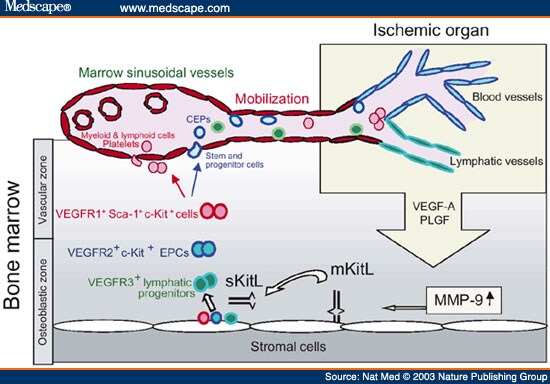
KTS: hyvä kuva!
http://img.medscape.com/fullsize/migrated/457/292/nm457292.fig2.jpg

http://journals.prous.com/journals/dnp/20051804/html/dn180257/images/francois_f2.gif

TULEHDUS merkkaa kriittisesti kardiovaskulaaristen tautien kehittymisessä.
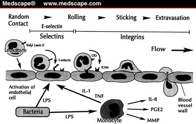
 Leukosyyttien infiltraatio vauriokohtiin vaatii niiden lähtemistä pois verestä ja migraatiota basaalikalvon läpi. Tällainen prosessi taas vaatii extrasellulaarisen matrixin (ECM ) remodelling-tapahtuman, hyvin kouriintuntuvan muokkauksen.
 Niinpä tässä ilmenee  tarvetta käyttää rheologisen alueen resurssimolekyyliä plasminogeenia Plg, joka on proteaasi, joka kiinittyy extrasellulaariseen matriksiin (ECM) ja kun se konvertoituu, muuttuu aktiiviin muotoon, plasmiiniksi (plm), hajoaa monta ECM-proteiinia. mikä degradaatio  kuuluu myös plasmiinin yhteen tehtävalueeseen.
http://img.medscape.com/fullsize/migrated/409/578/pharm2007.08.fig2.jpg

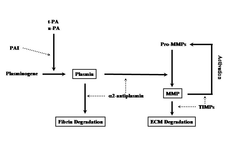
PLASMIINI pystyy aktivoimaan aivan suoraa MMP-järjestelmän.
 MMP entsyymiot  ovat matrixmetalloproteinaaseja, entsyymeitä, joita tunnetaan yli 20 erilaista.
Tässä työssään tutkijat käyttivät plasminogeeni-poistogeenista (KO) hiirtä (Plg-/-) , jotta he saisivat selville, mikä on PLASMINOGEENIN rooli tulehduksellisten leukosyyttien migroituessa.

Sitten koehiirille tehtiin peritoniittia vastaava tila ( vatsakalvontulehdustila). Ne hiiret, joilta puuttui plasminogeeni, osoittivat heikentynyttä makrofagien siirtymää ECM basaalikalvon läpi ja MMP-9 aktivaatio oli myös niillä alentunut. Mutta jos niille nyt annettiin MMP-9 injektio, tämä vaikutti makrofagitilanteen korjaantumiseen tässä koe-eläinmallissa.

Sitten he tutkivat toista tautimallia hiiressä, vatsa-aortan aneurysmamallia(AAA) . Jos näiltä hiiriltä nyt puuttui plasminogeeni ja sen takia niillä oli vähempi määrä makrofagi-infiltraatiota, ne olivat suojassa aortan aneurysman muodostumiselta. Mutta jos niille nyt annettiin aktiivia MMP-9 metalloproteinaasia, mikä edisti makrofagien paikalletuloa, niissä alkoi kehittyä vatsa-aortan aneurysma. ( Kts. MONOSYYTTI- MAKROFAGI linja!)

Näistä tiedoista tutkijat päättelivät, että PLASMINOGEENI (Plg) säätelee makrofagien vaeltamista tulehduskohtaan aktivoimalla metalloproteinaaseista MMP-9 tekijän.
MMP-9 taas puolestaan säätelee solujen ( esim monosyyttien)  kykyä migroitua ECM kalvon läpi. Sen takia tulehdusvasteitten säätelyssä saattaa Plg/MMP-9 tie olla kiinnostava lähtökohta kun koetetaan hallita vatsa-aortan aneurysman kehittymistilannetta

Plasmiinin osuus matrixmetalloproteinaasien päällekytkeytymiseen on uudehkoa tietoa.., TÄMÄ on asiasta ensimmäinen kaava jonka löysin vuosia sitten:
http://www.ophtalmo.ulg.ac.be/images/schemaplm-pln.jpg

Tämä on konservatiivinen tieto plasminogeenista:
http://www.geocities.com/omfsesan/plasminogen2.gif

Plasminogeenin aktivaatio: Alalolevaan kuvaan on merkattu MMP aktivaatio plasmiinin funktioksi. Tähän voisi vielä merkata  TAFI aktivaatiokin plasmiinin funktioksi .
http://www.iovs.org/misc/newClarkFig1.jpg

Tausta inhibitio PAI sen proangiogeenisyys ja antiangiogeenisyys  kuvana.
http://www.iovs.org/misc/newClarkFig2.jpg

22.5.2009 16:05