Etiketter

torsdag 4 januari 2018

Trombomoduliinin osuus MMP-järjestelmässä

JOS TM eli trombomoduliinimolekyyli puuttuu solupinnoista,   mitä sitten?
Tilannehan on mahdoton ihmiselle. 
http://atvb.ahajournals.org/content/36/4/728

 Lisääntyvän tiedon mukaan pre-eklampsia aiheutuu plasentalle ja endoteelille  välttämättömien tekijöiden  deprivaatiosta  ja siitä seuraa  istukan ja suonen pinnan, endoteelin,  toiminnan häiriö. Tässä viiteen artikkelista  osoitetaan, että  trombomoduliini mRNA:n  ja  vastaavan proteiinin ilmeneminen on alentunutta liveästi tai vaikeasti pre-eklamptisilla potilailla. ja tämä aleneminen korreloi negatiivisesti  odotavan äidin kehon painoindeksiin(BMI) ja diastolisessn paineeseen. Lisäksi trombomoduliini mRNA:n ilmenemä korreloi suoraan  plasentan matrixmetaalloproteinaasien  (MMP) ilmenemään ja jos istukan trombomoduliini  (TM) on  vähentynyttä, siitä seuraa  huonontunut  nukkasolujen elossapysyminen.

  • Increasing evidence suggests that preeclampsia is caused by deprivation of factors essential for the placenta and endothelium, with consequent placental and endothelial dysfunction. This study demonstrates that thrombomodulin mRNA and protein expression are decreased in placentas of mildly and severely preeclamptic patients, and that this decrease correlates negatively with maternal body mass index and diastolic blood pressure. Additionally, thrombomodulin mRNA expression correlates directly with placental expression of MMPs, and the decrease in placental thrombomodulin is accompanied by impaired villous cell survival. 


aPC aktivoi proMMP2 - linkki negatiivisen koagulaatioiojärjestelmän ja MMP järjestelmän välillä

Antikoagulaatiojärjestelmän ja MMP-järjestelmän väliset linkit:

Kogulaatiojärjestelmän suora linkki MMP-järjestelmään tiedetään fibrinolyysikaskadin   puolelta   siitä, että aktivoitunut plasmiini aktivoi sekventiaalisessa toiminnassa  myös MMP-järjestelmää yhteistyöhän (remodeling)
Koagulaatiojärjestelmä ja sen fysiologinen antikoagulaatiojärjestelmä ovat ajallisesti sekventiaalisia "overlapping ajallisesti"
Antikoagulaatiojärjestelmästäkin käsin tulee signaalia, joka aktivoi MMP-tekijää.
Alla oleva artikkeli  on vuodelta  2001.
Tämä antikoagulaatiojärjestelmä on sikäli hyvin mielenkiintoinen, koska ihminen pystyy vaikuttamaan sen hyvään kuntoon  käyttämällä K-vitamiinipitoista ravintoa. Nimittäin  C-proteiini on K-vitamiinista  riippuvainen, jotta se olisi toiminnallinen.
Hyytymisjärjstelmässä on usea peptidi K-vitamiinista  riippuva  (VKDP, Vitamin K Dependent peptides) mutta  niitä on myös  antikoagulaatiojärjestelmässä
S- proteiini ja S- proteiini ainakin.

Jos joudutaan käyttämään anti-K-vitamiinilääkitystä ( antikoagulanttia,  vaikuttuu sekä koagulaatio-  että  tämän fysiologinen antikoagulaatiojärjestelmän funktionaalisuus. Tämän  monivivahteisen järjestelmän  hyvään toimintaan voi vaikuttaa jo  lapsuudesta siten, että lapset ja nuoret totutetaan  aina päivittäin käyttämään  vihanneksia ja jonkin verran kasvisöljyjä, joissa on K1- vitamiinia. Kohtalaista liikuntaa  ja selalisia  ravinto-ohjeita kuten NNR  yleensä neuvoo. muutenkin neuvoo!

 Jos on  jo antikoagulanttilääkkeen käyttäjhä ja koko  koko  reologia  jo erilaisten pillerien varassa, on vaikea saada  normalisoitumista
  täysin palautumaan  alkuperäiseen kirjan kuvan kaavaan ilman  lääkkeiten antamia lisäapuja, mutta  on hyvä tietää, että  kun ei vielä ole riippuvainen
 antikoagulaatiolääkkeistä,  voi pitää omaa luonnollista antikoagulanttijärjestelmäänsä vireillä liikunnan  lisäksi   vihannes-
ja salaattiravinnolla, jota tulisi olla joka aterian joukossa vähäsen päivän mittaan. Esim salaatinlehti tavallisen välipalaleivän päällä.
Jos näitä pienia salaattiaterioita olisi päivittäin viiteen otteeseen hieman, se alkaisi vuosien varrella stabiloida myös   erään pahimman "villervallan" hyytymistekijöissä, siis vapaan kalsiumjonin, joka alkaisi asettua sille tarkoitettuihin neutraaleihin sijainteihin, kuten   luuston remodeling jrärjstelmään,  funktionaalisiin peptideihin , omiin aitioihinsa,  
pitämään  inaktiiviksi tarkoitetut muodot   inaktiiveina ja
solupinnat n neutraalikalvoina. Kalsiumjoni on hyytymisfaktori FIV.

Tässä yhteydessä, artikkelissa,  oletetaan että (kts kuva linkistä)

https://openi.nlm.nih.gov/detailedresult.php?img=PMC538749_1471-2377-4-21-1&req=4

C-proteiini on toiminnallinen, siis saanut posttranslationaalisen kalsiumkatteensa ja pystynyt aktivoitumaan normaaliin tapaan aPC muotoon ja  osallistumaan  antikoagulaatioon ( trombiinin ja trombomoduulin aktivoimana  aPC- molekyylinä). 

Tässä  artikkelissa kerrotaan, että aPC-proteiinilla on  mahdollisuus antaa signaalia ehkä  jo edeltä MMP-järjstelmään ennen kuin  plasminogeeni on ehtinyt  aktivoitua plasmiiniksi (?)  tai ehkä plasmiiniaktivaatiossa on jokin viive tai vikatie menossa, jolloin  juuri MMP2 painottuu. 


LÄHDE:  FEBS Lett. 2001 Apr 6;494(1-2):129-32.
Thrombin-thrombomodulin activation of protein C facilitates the activation of progelatinase A.
Pekovich SR1, Bock PE, Hoover RL.


TIIVISTELMÄ  MMP-progelatinaasi A- (proMMP-2)  aktivaatio on kiinnostava, koska  sitä ilmenee  sairaustiloissa syövässä ja ateroskleroosissa.
  • Abstract
  • The activation of the matrix metalloproteinase progelatinase A (MMP-2) has been of keen interest because an increase in MMP-2 activity has been implicated in disease states such as cancer and atherosclerosis.
 MMP-2 aktivaatio tapahtuu kahdessa  aihessa spesifisten solujen pinnalla.  ensimmäisessää vaiheessa  MT-MMP- tyyppinen  proteaasi  pilkkooo  MMP-2 välituotetta Toisessa pilkkoutumisessa  ja välituotteen aktivaatiossa   toimii  autokatalyysi, näin on ajateltu. 

  •  Activation of MMP-2 occurs on the surface of specific cell types in two steps.  In the first step, primary cleavage of MMP-2 by a membrane-type matrix metalloproteinase (MT-MMP)  generates an intermediate. A secondary cleavage and activation of the intermediate is thought to occur autocatalytically.

Aiemmat tutkimukset ovat osoittaneet, että trombiini voi myös aktivoida progelatinaasi A :ta endoteelisolujen läsnäollessa-  Tutkijat osoittavat, että tämä soluista riippuva mekanimsi, jossa MMP-2 aktivoituu, toimii myös THP-1 soluissa ja se käsittää  trombiinin sitoutumista trombomoduliin TM), jota on solun pinnalla ja  C-proteiinin kanssa  tästä muodostuu aktivoitunut C-proteiini aPC. , joka on antikoagulantti.

  • Previous studies have shown that thrombin can also activate progelatinase A in the presence of endothelial cells. We show that this cell-dependent mechanism of MMP-2 activation also occurs with THP-1 cells and involves binding of thrombin to thrombomodulin present on the cell surface and generation of the anti-coagulant protein, activated protein C.
Sitten tutkijat osoittivat, että  aktivoitunut C-proteiini on suoraan  aktivoimassa ja pilkkomassa gelatinaasiA-välituotetta.  Tämä työ osaltaan  antaa uusia mekanistisia oivalluksia MMP-2-aktivaatiosta ja  tarjoaa uuden linkin MMP-aktivaation ja antikoagualation kesken.
  •  We demonstrate that activated protein C is directly responsible for activation and cleavage of the gelatinase A intermediate. This work contributesnew mechanistic insights into the activation of MMP-2 and provides a novel link between matrix metalloproteinase activation and anti-coagulation.,

Onkolyyttinen tuhkarokkovirus syöpäterapiassa

https://www.ncbi.nlm.nih.gov/pmc/articles/PMC5086626/
 2016

Measles to the Rescue: A Review of Oncolytic Measles Virus

Richard K. Plemper, Academic Editor
Abstract
Oncolytic virotherapeutic agents are likely to become serious contenders in cancer treatment. The vaccine strain of measles virus is an agent with an impressive range of oncolytic activity in pre-clinical trials with increasing evidence of safety and efficacy in early clinical trials. This paramyxovirus vaccine has a proven safety record and is amenable to careful genetic modification in the laboratory. 

Overexpression of the measles virus (MV) receptor CD46 in many tumour cells may direct the virus to preferentially enter transformed cells and there is increasing awareness of the importance of nectin-4 and signaling lymphocytic activation molecule (SLAM) in oncolysis. Successful attempts to retarget MV by inserting genes for tumour-specific ligands to antigens such as carcinoembryonic antigen (CEA), CD20, CD38, and by engineering the virus to express synthetic microRNA targeting sequences, and “blinding” the virus to the natural viral receptors are exciting measures to increase viral specificity and enhance the oncolytic effect. 

Sodium iodine symporter (NIS) can also be expressed by MV, which enables in vivo tracking of MV infection. Radiovirotherapy using MV-NIS, chemo-virotherapy to convert prodrugs to their toxic metabolites, and immune-virotherapy including incorporating antibodies against immune checkpoint inhibitors can also increase the oncolytic potential. 

 Anti-viral host immune responses are a recognized barrier to the success of MV, and approaches such as transporting MV to the tumour sites by carrier cells, are showing promise. MV Clinical trials are producing encouraging preliminary results in ovarian cancer, myeloma and cutaneous non-Hodgkin lymphoma, and the outcome of currently open trials in glioblastoma multiforme, mesothelioma and squamous cell carcinoma are eagerly anticipated.
Keywords: measles virus, oncolytic, virotherapy

Tuhkarokkovirus ja MMP-järjestelmä. pohdittavia artikkeleita

Haku PubMed Measles and MMP, 6 vastausta:

Search results

Items: 6

1.
Mühlebach MD, Schaser T, Zimmermann M, Armeanu S, Hanschmann KM, Cattaneo R, Bitzer M, Lauer UM, Cichutek K, Buchholz CJ.
Cancer Res. 2010 Oct 1;70(19):7620-9. doi: 10.1158/0008-5472.CAN-09-4650. Epub 2010 Sep 21.

Abstract

Primary and secondary cancers of the liver are a significant health problem with limited treatment options. We sought here to develop an oncolytic measles virus (MV) preferentially activated in liver tumor tissue, thus reducing infection and destruction of healthy tissue. We documented that in primary tumor tissue, urokinase-type plasminogen activator and especially matrix metalloproteinase-2 (MMP-2) are significantly more active than in adjacent nontumorous tissue. We then generated variants of the MV fusion protein by inserting different MMP substrate motifs at the protease cleavage site and identified the motif PQGLYA as the most efficient cleavage site as determined by syncytia formation on protease-positive tumor cells. The corresponding MMP-activatable oncolytic MV-MMPA1 virus was rescued and shown to be strongly restricted on primary human hepatocytes and healthy human liver tissue, while remaining as effective as the parental MV in the tumor tissue sections. Our findings underline the clinical potency of the MMP activation concept as a strategy to generate safer oncolytic viruses for the treatment of primary and secondary cancers of the liver.
Free Article
2.
Beineke A, Puff C, Seehusen F, Baumgärtner W.
Vet Immunol Immunopathol. 2009 Jan 15;127(1-2):1-18. doi: 10.1016/j.vetimm.2008.09.023. Epub 2008 Oct 4. Review.
PMID:
19019458
3.
Ichiyama T, Matsushige T, Siba P, Suarkia D, Takasu T, Miki K, Furukawa S.
J Infect. 2008 May;56(5):376-80. doi: 10.1016/j.jinf.2008.02.014. Epub 2008 Apr 18.

Abstract

OBJECTIVES:
To investigate the brain inflammation and damage in subacute sclerosing panencephalitis (SSPE), the cerebrospinal fluid (CSF) concentrations of matrix metalloproteinase-9 (MMP-9) and tissue inhibitor of metalloproteinase-1 (TIMP-1) were determined in SSPE patients.
METHODS:
CSF MMP-9 and TIMP-1 levels were measured in 23 patients with SSPE in Papua New Guinea by ELISA.
RESULTS:
CSF MMP-9 levels and MMP-9/TIMP-1 ratios of SSPE patients were significantly higher than controls (p<0 .001="" and="" between="" controls.="" csf="" differences="" in="" levels="" no="" p="" patients="" respectively="" significant="" sspe="" there="" timp-1="" were="">CONCLUSIONS:
Previous studies suggested that CSF MMP-9 levels reflect inflammatory damage to the brain. Our findings suggest that the MMP-9 level in CSF is an indicator of inflammatory damage to the brain in SSPE.
4.
Ichiyama T, Siba P, Suarkia D, Takasu T, Miki K, Kira R, Kusuhara K, Hara T, Toyama J, Furukawa S.
J Neurol Sci. 2007 Jan 15;252(1):45-8. Epub 2006 Nov 22.

Abstract

We determined the relationship between the serum concentrations of matrix metalloproteinase-9 (MMP-9) and tissue inhibitors of metalloproteinases 1 (TIMP-1) in 33 patients with subacute sclerosing panencephalitis (SSPE) to investigate the function of the blood-brain-barrier (BBB) in SSPE. Serum MMP-9 and TIMP-1 levels were measured by ELISA. Serum MMP-9 levels and MMP-9/TIMP-1 ratios of SSPE patients in Papua New Guinea (n = 24), and those in Japan (n = 9) were significantly higher than the each control (MMP-9, p = 0.0390, and p = 0.0023, respectively; MMP-9/TIMP-1, p = 0.0319, and p = 0.0009, respectively). Serum MMP-9 levels and MMP-9/TIMP-1 ratios of SSPE patients with Jabbour stage III (n = 13) were significantly higher than those with Jabbour stage II (n = 18) (p = 0.003, and p = 0.0412, respectively). There were no significant differences of serum TIMP-1 levels between the SSPE patients and controls. High serum MMP-9 and MMP-9/TIMP-1 levels will promote brain invasion through the BBB by immunocompetent cells in the blood. Our findings suggest that the balance of serum MMP-9 and TIMP-1 levels modulate the inflammatory cascade of SSPE.
5.
Springfeld C, von Messling V, Frenzke M, Ungerechts G, Buchholz CJ, Cattaneo R.
Cancer Res. 2006 Aug 1;66(15):7694-700.

Abstract

Cancer cells secrete matrix metalloproteinases (MMP) that degrade the extracellular matrix and are responsible for some hallmarks of malignant cancer. Many viruses, including a few currently used in oncolytic virotherapy clinical trials, depend on intracellular proteases to process their proteins and activate their particles. We show here for measles virus (MV) that particle activation can be made dependent of proteases secreted by cancer cells. The MV depends on the intracellular protease furin to process and activate its envelope fusion (F) protein. To make F protein activation cancer cell specific, we introduced hexameric sequences recognized by an MMP and identified the mutant proteins most effective in fusing MMP-expressing human fibrosarcoma cells (HT1080). We showed that an MMP inhibitor interferes with syncytia formation elicited by mutant F proteins and confirmed MMP-dependent cleavage by Edman degradation sequence analysis. We generated recombinant MVs expressing the modified F proteins in place of furin-activated F. These viruses spread only in cells secreting MMP. In nude mice, an MMP-activated MV retarded HT1080 xenograft growth as efficiently as the furin-activated MV vaccine strain. In MV-susceptible mice, the furin-activated virus caused lethal encephalitis upon intracerebral inoculation, whereas the MMP-activated did not. Thus, MV particle activation can be made dependent of proteases secreted by cancer cells, enhancing safety. This study opens the perspective of combining targeting at the particle activation, receptor recognition, and selective replication levels to improve the therapeutic index of MV and other viruses in ongoing clinical trials of oncolysis.
6.
Agafonov AP, Kameneva SN, Agafonova OA, Neverov AA, Ignat'ev GM.
Vestn Ross Akad Med Nauk. 2004;(8):3-6. Russian.

Abstract

The level of specific antibodies to viruses of measles, parotitis, type-6 herpes, Epstein-Barr, tick-borne encephalitis and Borrelia burgdorferi as well as presence of genetic samples and antigens of the above infectious antigens were studied in patients with multiple sclerosis (MS). The cytokines Th1 and Th2 parameters were investigated in blood serum of patients at different MS stages. The titer of antibodies to measles virus was noted to be increasing in MS patients with age and disease aggravation. The level of antibodies to any of the studied infectious agents, except for the type-6 herpes virus, was not dynamically changing for as long as 9 months. The viral genetic samples (measles RNA) were detected just once in 2 patients; the detection time coincided in both cases with MS aggravation. The cytokines dynamics failed to correlate with MS aggravation or exacerbation while the total index of all studied cytokines was decreased. A high MMPw 9 content in blood serum correlated with MS exacerbation in 1 patient.

onsdag 3 januari 2018

Pravastatiini vaimentaa MMP mRNA ja proteiinisynteesiä

https://link.springer.com/article/10.1007/s10195-012-0200-4

Journal of Orthopaedics and Traumatology
, Volume 13, Issue 3, pp 119–123 | Cite as

Pravastatin suppresses matrix metalloproteinase expression and activity in human articular chondrocytes stimulated by interleukin-1β

MMP-järjestelmä voi aktivoitua katepsiinien taholta

https://www.ncbi.nlm.nih.gov/pmc/articles/PMC3856311/

World J Biol Chem. 2013 Nov 26; 4(4): 91–101.
Published online 2013 Nov 26. doi:  10.4331/wjbc.v4.i4.91
PMCID: PMC3856311

Cathepsins mediate tumor metastasis

Abstract
Cathepsins are highly expressed in various human cancers, associated with tumor metastasis. It is superfamily, concluding A, B, C, D, E, F, G, H, L, K, O, S, V, and W family members. As a group of lysosomal proteinases or endopeptidases, each member has a different function, playing different roles in distinct tumorigenic processes such as proliferation, angiogenesis, metastasis, and invasion. Cathepsins belong to a diverse number of enzyme subtypes, including cysteine proteases, serine proteases and aspartic proteases. The contribution of cathepsins to invasion in human cancers is well documented, although the precise mechanisms by which cathepsins exert their effects are still not clear. In the present review, the role of cathepsin family members in cancer is discussed.
Keywords: Cathepsin, Tumor, Metastasis, Mechanism


Lisätietoa matrixmetalloproteinaaseista

Tämä on sitaatti netistä ja  viittaa kirjaan.   Suomennan kappale kappaleelta

Learn more about Matrix metalloproteinase

Matrix metalloproteinases (MMPs) are a family of highly homologous protein degrading zinc dependent endopeptidases that break down components of the extracellular matrix.
From: Advances in Pharmacology, 2012http://www.sciencedirect.com/topics/medicine-and-dentistry/matrix-metalloproteinase


1. Matrix Metalloproteinase 3/Stromelysin 1


Hideaki Nagase, in Handbook of Proteolytic Enzymes (Third Edition), 2013
Distinguishing Features
(kts.  suomennos 3.1. 2018 : Lisätieto stromelysiineistä ST-1, -2)

2. Matrix Metalloproteinase-11/Stromelysin 3

Marie-Christine Rio, in Handbook of Proteolytic Enzymes (Third Edition), 2013
Name and History
(kts.  suomennos 3.1. 2017 : Lisätieto stromelysiineistä, ST-3)

3. Molecular Biology of Head and Neck Cancer

David K. Lam, Brian L. Schmidt, in Current Therapy In Oral and Maxillofacial Surgery, 2012

Matrix Metalloproteinases

Matrix metalloproteinases (MMPs), or zinc metalloenzymes involved in extracellular matrix remodeling, consist of the collagenases, gelatinases, stromelysins, stromelysin-like MMPs, membrane-type MMPs,
 and new MMPs. Malignant cells may use MMPs to dissolve the basement membrane and degrade interstitial stroma  to facilitate invasion and metastasis. MMP-2 expression is higher in cell nests of metastatic tumors than in those of non-metastatic tumors in oral cancer, thus suggesting that MMP-2 is a marker predictive of metastasis.
142,143
MMP-3, MMP-10, and MMP-11(stromelysiinit)  have also been implicated in the progression of oral cancer.144,145

Read full chapter

4. Matrix Metalloproteinase 23


Duanqing Pei, in Handbook of Proteolytic Enzymes (Third Edition), 2013
Name and History

Matrix metalloproteinase 23 (MMP-23) is the first type II transmembrane matrix metalloproteinase described
 in the matrix metalloproteinase family [1]. It has been isolated from multiple species including human,
 mouse and rat [2–5]. The first evidence of its sequence was reported by Gururajan and colleagues [2]
 describing the identification of two MMPs genes named MMP-21 and MMP-22 in duplicate within human chromosome
1p36.3 region frequently implicated in tumor suppression. However, these numbers had previously been assigned
to other MMPs. Subsequently, a human cDNA was isolated and named MMP-23; this had almost identical sequence
to the reported MMP21/22 [5]. The mouse cDNA was isolated and named CA-MMP (Cysteine Array MMP) based on
 structural features [4]. The same gene has also been identified in the female reproductive systems of human
and rat and named MIFR/Femalysin accordingly [3]. The name MMP-23 has been officially recognized as the
serial designation within the MMP family [6].

Read full chapter

5. Viruses Responsible for Hemorrhagic Fevers

Vivian Vasconcelos Costa***†, ... Mauro Martins Teixeira**, in Vascular Responses to Pathogens, 2016
Matrix Metalloproteinases

Matrix metalloproteinases (MMPs) or matrixins are proteases that break extracellular matrix (ECM)
and proteins that lay out the ECM, while the tissue inhibitor of matrix metalloproteinases (TIMPs)
 block the activity of these MMPs and prevent the destruction of ECM. Thus, the balance between MMPs and TIMPs
 is critical for the eventual ECM remodeling in the tissue 122 and these molecules have shown important
roles in many physiological and pathological conditions.123 In this context, Luplertlop et al. showed
that DENV infection of immature dendritic cells (iDCs) led to overproduction of soluble matrix metalloproteinases MMP-9 and MMP-2
and that these enzymes were capable of enhancing endothelial permeability of HUVEC monolayer cells in vitro and
 in a model of vascular permeability in vivo.84 Of note, MMP-9 production was enhanced in patients with DENV infection
 and was associated with severity of disease.85 Indeed, elevated levels of MMPs and their correlation with
severe disease do not appear to be exclusive to DENV infections. Genome-wide host expression patterns
 in spleen or blood samples from mice and nonhuman primates, respectively, infected with Ebola virus
revealed a positive correlation between the dysregulated inflammatory responses associated with induction
 of specific MMPs and the lethal outcome of Ebola.86,87 Interestingly, Aydin et al. showed that during the
acute phase of disease, severe CCHF-infected patients presented higher serum levels of the tissue inhibitor
 of MMPs (TIMP-1) than nonsevere patients. Indeed, severe patients also revealed enhanced levels of MMP-1, 7, 9, 10
during the convalescence phase, suggesting that the imbalance between these molecules plays a role
 in the pathogenesis of CCHF.88

Read full chapter

6. Angiogenesis and Anti-Angiogenesis Strategies in Cancer


Shaker A. Mousa, Paul J. Davis, in Anti-Angiogenesis Strategies in Cancer Therapeutics, 2017
Matrix Metalloproteinases

Matrix metalloproteinases (MMPs) are zinc-dependent endopeptidases that degrade ECM proteins—collagen, laminin,
 and fibronectin—during the process of cancer invasion and metastasis. MMPs are produced primarily by
 reactive stromal and inflammatory cells surrounding tumors rather than by cancer cells.
 Hydroxamic acid-derived inhibitors of MMPs are orally bioavailable. Limited efficacy of
this class of agents in clinical trials against a variety of solid tumors prompted termination
 of clinical trials [33,34]. MMPs are also essential to angiogenesis, preparing the tumor microenvironment
for the expansion of tumor blood supply.

Read full chapter

7. Vascular Smooth Muscle


Lula L. Hilenski, Kathy K. Griendling, in Vascular Medicine: A Companion to Braunwald's Heart Disease (Second Edition), 2013
Matrix metalloproteinases and tissue inhibitors of matrix metalloproteinases

Matrix metalloproteinases are zinc-containing enzymes that, along with extracellular proteases
 in the plasminogen activation system, induce remodeling of VSMC cell-matrix and cell-cell interactions
(reviewed in92–94) and release ECM-bound growth factors, cytokines, and proteolyzed ECM fragments,
 or “matrikines,” with cytokine-like properties into the ECM. Members of the MMP family found
 in vascular tissues (listed in Ref. 95) include interstitial collagenases, basement membrane gelatinases,
stromelysins, matrilysins, and membrane type (MT)-MMPs and metalloelastase (see Chapter 4).
 In the vascular wall, production of pro-MMP-2, MMP-14, and TIMP-1 and -2 is constitutive,96
while other MMPs can be induced by inflammatory cytokines (interleukin [IL]-1 and -4 and tumor
necrosis factor α [TNF-α]), hemodynamics, vessel injury, and ROS.93 In addition, MMPs can act
synergistically with growth factors such as PDGF and FGF-2.

Matrix metalloproteinase induced remodeling of basement membrane components laminin,
 polymerized type IV collagen, and HSPGs promotes a VSMC migratory phenotype.
 In addition, MMP cleavage and shedding of non-matrix substrates—in particular,
 adherens junction cadherins—act to remove physical constraints on cell movement.93
Furthermore, ECM remodeling enables integrin signaling from the cell surface to focal adhesions,
 modulating cell cycle components cyclin D1 and p21/p27 CDKIs.96

In vascular remodeling, MMP activities are tightly regulated at several levels:
transcriptional level, activation of pro-forms, interaction with specific ECM components,
and inhibition by TIMPs. Modulation of MMP activity is evident in VSMC migration and neointima formation
after injury, plaque destabilization in atherosclerosis, aneurysm formation, hypertension, and coronary
restenosis.95
In atherosclerosis, MMPs have potential either to promote plaque instability, as in
advanced plaques of hypercholesterolemia models, or to stabilize plaques by increasing VSMC migration/proliferation.
Up-regulation of MMPs in VSMCs may contribute to aneurysm formation.3

Read full chapter

8. Neuroprotection: Where Are We Going?


Gary H. Danton MD, PhD, W. Dalton Dietrich PhD, in From Neuroscience To Neurology, 2005
Matrix Metalloproteinases (Table 14.3)

Matrix metalloproteinases (MMPs) are angiogenic enzymes that break down components of the
extracellular matrix. Although angiogenesis seems like a potential benefit,
MMPs may enhance BBB breakdown after stroke and increase inflammation. MMP inhibitors
such as batimistat (BB-94) and KB-R7785 decreased infarct volume in mice following permanent
focal ischemia (Asahi et al., 2000; Jiang et al., 2001). BB-1101 reduced edema 24 hours after
 focal ischemia in rats and reduced hemorrhage in both rats and rabbits (Rosenberg et al., 1998;
 Lapchak et al., 2000; Sumii et al., 2002). MMP

inhibitors have been evaluated in patients for their antiangiogenesis properties in cancer treatment
and are well tolerated in patients (Hidalgo and Eckhardt, 2001).

Read full chapter

9. Matrix Metalloproteinases in Biologic Samples


Gregory T. Jones1, in Advances in Clinical Chemistry, 2014
Matrix metalloproteinases (MMPs) are a subfamily of the metzincin superfamily
 of endogenous proteinases [1–3]. MMPs typically consist of a propeptide domain,
 a catalytic metalloproteinase domain, a linker (hinge region) peptide, and a hemopexin domain [1].
Common structural features consist of a zinc-binding motif (HEXXHXXGXXH) within the catalytic domain
 and a cysteine switch motif (PRCGXPD) within the propeptide domain [4] (Fig. 7.1).
The binding of these domains to the catalytic zinc ion keeps the proenzyme in an inactive state
by preventing water molecule binding, required for catalysis, to the zinc ion [1].
Understanding the nature of these activation states is key to interpreting any methodological
technique that attempts to measure MMP activity.

Read full chapter

10. Diagnostic Techniques in Ocular Surface Disease


Bennie H. Jeng, in Ocular Surface Disease: Cornea, Conjunctiva and Tear Film, 2013
Rapid Testing For Inflammatory Markers

Matrix metalloproteinase 9 (MMP-9) plays a critical role in wound healing
and inflammation, and is primarily responsible for the pathologic alterations
to the ocular surface in various conditions.36
MMP-9 has been demonstrated to be significantly elevated in the tears of patients
with blepharitis, allergic eye disease, dry eye disease, and conjunctivochalasis.37,38
The ability to test for MMP-9 in the tear film may prove to be an important tool to help
in the diagnosis of ocular surface disease. Recently, a commercially available
 point-of-care test, RPS InflammaDry Detector (RPS, Inc, Sarasota, FL, USA) offers
 an easy-to-administer and rapid turn-around test (10 minutes) for measuring MMP-9 levels in the tear film.

Read full chapter

Lisätieto stromelysiineistä ST-1, -2, -3.

(1)   MMP-3 , toiselta nimeltä stromelysiini 1, ST-1 

MMP-10, ST-2. 

 Matrix Metalloproteinase 3/Stromelysin 1 

Lähde: Hideaki Nagase, in Handbook of Proteolytic Enzymes (Third Edition), 2013

Erottavia piirteiä
MMP-3:lla (stromelysiini 1:llä)  ja MMP-1:lla  (interstitiaalisella kollagenaasilla) on sama molekyylimassa ja niitä erittyy samoista a soluista. MMP-3 eroaa MMP-1:stä  siinä että siltä puuttuu kyky pilkkoa kollageenityypeistä I ja II niiden kolmoishelixrakennetta sisältäviä kohtia.
Stromelysiini 1 (MMP-3)  ja stromelysiini 2 (MMP-10) ovat 78%:sti identtisiä  aminohappojärjestykseltään ja niiden entsyymiominaisuudet ovat samat, mutta pH-optimit ovat erilaisia. ST- 1 toimii optimaalisisti pH5.5-6.0 alueella ( happamassa miljöössä) ja ST-2  pH alueella 7.5- 8.0 eli emäksisessä miljöössä.

  • Distinguishing Features
  • Stromelysin 1 (MMP-3) and interstitial collagenase (MMP-1) have similar molecular masses and they  are often secreted from the same cells. MMP-3 can be distinguished from MMP-1 (Chapter 152)  by its failure to cleave triple helical regions of types I and II collagen. Stromelysin 1 and stromelysin 2 (MMP-10) (Chapter 159) share 78% identity in amino acid sequence and have similar enzymic properties but the pH optimum of stromelysin 1 is around pH 5.5–6.0, whereas that of stromelysin 2 is around pH 7.5–8.0 [19].
 Neutrofiilien elastaasi ei  aktivoi pro-stromelysiini-2:ta, vaikka se aktivoi pro-stromelysiini 1:n. näiden kahden stromelysiinin ilmeneminenkin soluissa on erilaista:  Stromelysiini-1 ilmenee  helposti mesenkymaalisissa soluissa, kun taas stromelysiini-2  ilmenee keratinosyyteissä tietyistä stimuluksista.
Stromelysiini-3 (ST-3) on  näistä  molemmista muista  stromelysiineistä rakenteellisesti ja entsyymiominaisuuksiltaan loitompi ( Siitä kerrotaan erikseen)

  •  Pro-stromelysin 2 is not activated by neutrophil elastase (Chapter 587) while prostromelysin 1 is.  The cellular expression of the two stromelysins are different: stromelysin 1 is readily expressed  in mesenchymal cells whereas stromelysin 2 is expressed in keratinocytes under certain stimuli [74,75]. Stromelysin 3 (Chapter 160) is more distantly related to these two stromelysins in structure and enzymic properties. 

 On kuvattu polyklonaalisia vasta-aineita  näille entsyymeille.  Kaniinin polyklonaalinen vasta-aine  ihmisen stromelysiini 1:tä vastaan ei reagoi ihmisen stromelysiini 2:ta vastaan.  On olemassa  ELISA-kit ihmisen MMP-3:lle.

  • Polyclonal antibodies against human [76,77], rabbit [78], cattle [79] and rat [80],and monoclonal antibodies against human [81] and rabbit [82] enzymes have been described.  Rabbit polyclonal antibody against human stromelysin 1 [76] fails to react with stromelysin 2 [75]. A commercial ELISA kit for human MMP-3 is available from GE Healthcare, Abcam, R&D System; antibodies reactive with human, pig, sheep, mouse and rat MMP-3 from Abcam.

(2) MMP-11 eli stromelysiini 3  (ST-3)

Stromelysiini 3 cDNA  kloonattiin ensi kertaa  kun tehtiin differentiaalidiagnostiikkaa malignien ja benignien rintasyöpänäytteiden suhteen. Silloin  tunnistettiin stromelysiini 3  stromelysiini-aölaperheen kolmanneksi jäseneksi  metalloproteinaasien  perheessä, koska sillä oli jotain samankaltaisuutta  ST_1 ja ST-2:n kanssa
Vaikka se täten alunperin merkattiin ST3:ksi,  sen sekvenssi on aika poikkeava ST-1 ja ST-2 sekvensseistä. Täten nimitys "stromelysiini 3" ei  heijasta mitään funktionaalisia kaltaisuuksia toisten stromelysiinien kanssa.
  • LÄHDE: Marie-Christine Rio, in Handbook of Proteolytic Enzymes (Third Edition), 2013: Matrix Metalloproteinase-11/Stromelysin 3 Name and History
  • The stromelysin 3 (ST3) cDNA was first cloned by differential screening between malignant and benign human breast tumor samples [1]. ST3 was initially recognized as the third memberof the stromelysin subfamily within the matrix metalloproteinase (MMP) family on the basis of its similarity with the stromelysin 1 (ST1) and stromelysin 2 (ST2). Despite the original designation as ST3, its sequence is quite divergent from the ST1 and ST2and it has distinct mechanism of activation and proteolytic activity [2–4]. Thus, the name ST3 does not reflect any functional similarities with other stromelysins.
 Kun oli keksitty MMP- sekvenssi saatiin ST3:lle   geneerisesti tarkempi kuvaus ja se merkattiin  matrixmetalloproteinaasiksi numero  11 (MMP-11) . Muiden matrixproteiinien tavoin ST-3 osallistuu extrasellulaarisen matrixin uudelleenmuokkaustapahtumiin (remodelling) , jota  esiintyy kehityksen aikana, kudosten surkastuessa, haavan paranemisessa, tuumorin leviämissä ja etäispesäkkeiden muodostuksessa. Kuitenkin kirjallisuudesta päätellen ST3 näyttää olevan  ainutlaatuinen jäsen MMP perheessä ja sillä on  omat tyyppipiirteensä ja funktionsa.

  •  A more generic designation based on the sequence of MMP discovery has assigned ST3 the serial number 11, i.e. matrix metalloproteinase 11. As the other MMPs, ST3 is involved during extracellular matrix (ECM) remodeling events such as development, tissue involution, wound healing, tumor invasion and metastasis [5–8]. However, from literature, ST3 appears to be a unique member of the MMP family exhibiting peculiar features and function (reviewed in Murphy et al. [9]).

tisdag 2 januari 2018

Pro-MMP voi aktivoitua oksidatiivisesta stressistä

Peroxynitriitti  indusoi MMP aktivaation.

http://www.jbc.org/content/276/31/29596.short

Activation of Matrix Metalloproteinases by Peroxynitrite-induced Protein S-Glutathiolation via Disulfide S-Oxide Formation*

    Tatsuya Okamoto‡,§, Takaaki Akaike‡¶, Tomohiro Sawa‡, Yoichi Miyamoto‡, Albert van der Vliet§ and Hiroshi Maeda‡

+
Author Affiliations

    From the ‡Department of Microbiology,
 Kumamoto University School of Medicine, Kumamoto 860-0811,
Japan and the §Division of Pulmonary/Critical Care Medicine,
Department of Internal Medicine, School of Medicine,
 University of California, Davis, California 95616

TIIVISTELMÄ
Abstract

Oxidatiivinen stressi voi aiheuttaa kudosvauriota aktivoimalla matrixmetaalloproteinaasien (MMP)  esimuotoja (proMMP) . 
  • Oxidative stress may cause tissue injury through activation of the precursors of matrix metalloproteinase (proMMPs).
 Tässä tutkimuksessa havainnoitiin  glutationista riippuvaa pro-MMP-aktivaatiota, jota aiheutti peroxynitriitti. Se on voimakas oksidoiva agenssi, jota muodostuu  tulehduksellisissa prosesseissa. Peroxynitriitt aktivoi  kaikki kolme tutkittua  puhdistettua humaania  pro MMP-lajia (proMMP-1, proMMP-8 ja proMMP-9)  samanvahvuisissa GSH -pitoisuuksissa.
  •  In this study, we observed glutathione (GSH)-dependent proMMP activation induced by peroxynitrite,  a potent oxidizing agent formed during inflammatory processes. Peroxynitrite strongly activated all  three types of purified human proMMPs (proMMP-1, -8, and -9) in the presence of similar concentrations of GSH.
 Peroxynitriitin ja GSH:n välisistä reaktiotuotteista vain  yksi laji, S-nitroglutationi (GSNO2)  aiheutti proMMP-aktivaatiot. ProMMP-aktivaatiossa tapahtui  laaja-alainen S-glutatiolaatio  proMMP-proteiinissa kun aktivoitiin  tätä edeltäjämolekyyliä peroxynitriitillä ja GSH:lla.  Reaktiota seurattiin  radioisotoopein merkattujen GSH-molekyylien avulla . Käytettiin joko [35S]GSH tai [3H]GSH merkkauksia. Huomattavasta S-glutatiolaatiosta oli  näyttöä vielä ditiotreitoli ja proteiinidenaturaatiokäsittelyn jälkeenkin. Kuitenkin  oli viitettä siitä, että osa S-glutatiolaatiosta ei tapahtunut yksinkertaisen  disulfidimuodostuksen kautta.
MALDI-TOF -massaspektrometrisesti osoitettiin, ettei ainoastaan peroxynitriitti + GSH tuottanut ditiotreitoliresistenttiä S-glutatiolaatiota    synteettiseen peptidiin PRCGVPD ( joka on tunnettu  proMMP:n autoinhibitorinen domaani, jossa on konservoitunut cysteiinin sisältävä jakso)- sillä myös  syntetisoitunut GSNO2 tuotti ditiothreitolresistenssin. 
Oletetaan PRCGVPDS- glutatiolaation  tapahtuvan  glutationi disulfidi S-oxidin muodostumisen kautta  (GS(O)SR). Nämä tulokset osoittavat  ainutlaatuisen mekanimsin, jolla  proMMP aktivoituu oxidatiivisesti ja kudos  kärsii tulehduksen aiheuttamasta oksidatiivissta vauriosta.

  • Of the potential reaction products between peroxynitrite and GSH, only S-nitroglutathione (GSNO2) caused  proMMP activation. ExtensiveS-glutathiolation of the proMMP protein occurred during activation of proMMP by peroxynitrite and GSH, as shown by radiolabeling studies with [35S]GSH or [3H]GSH. Evidence of appreciable S-glutathiolation persisted even after dithiothreitol and protein-denaturing treatment,  however, suggesting that some S-glutathiolation did not occur through formation of simple mixed disulfide. Matrix-assisted laser-desorption ionization-time-of-flight mass spectrometry  indicated that not only peroxynitrite plus GSH but also synthetic GSNO2 produced dithiothreitol-resistant S-glutathiolation of the synthetic peptide PRCGVPD, which is a well conserved Cys-containing sequence  of the propeptide autoinhibitory domain of proMMPs. PRCGVPDS-glutathiolation is presumed to be formed through glutathione disulfide S-oxide (GS(O)SR), based on them/z 1064. Our results illustrate a unique mechanism of oxidative proMMP activation and oxidative tissue injury during inflammation.
LYHENNYKSIÄ:
Abbreviations:

ECM
    extracellular matrix , Extrasellularinen matriksi
MMP
    matrix metalloproteinase, matrixmetalloproteinaasi
proMMP
    precursors of MMP, MMP- edeltäjämolekyyli
IPF
    idiopathic pulmonary fibrosis, keuhkofibroosi
PCMB
    p-chloromercuribenzoate
NEM
    N-ethylmaleimide
PAGE
    polyacrylamide gel electrophoresis
HPLC
    high performance liquid chromatography
NAC
    N-acetyl l-cysteine
BSA
    bovine serum albumin
GSNO
    S-nitrosoglutathione
GSNO2

    S-nitroglutathione
MALDI-TOF MS
    matrix-assisted laser-desorption ionization-time-of-flight mass spectrometry
CBB
    Coomassie Brilliant Blue
DTT
    dithiothreitol


    Received March 19, 2001.
    Revision received May 3, 2001.

    The American Society for Biochemistry and Molecular Biology, Inc.


MMP perheen biologinen rooli ja kriittinen tasapaino

http://erj.ersjournals.com/content/38/1/191
Matrixmetalloproteinaaseilla on biologinen tehtävänsä ja  tasapaino tässä järjestelmässä on kriittistä ihmsen hyvinvoinnille.  Tämä järjestelmä on kiintesäti liittynyt kehon  rheologiseen tasapainoon sikäli että veressä olevien tekijöiden kautta taphtuu MMP aktivaatio kaikkien taiteen sääntöjen mukaan  eduksi  kehon  remodelling tarpeelle esim hyytymien pilkkoutumisen yhteydessä ja haavojen korjaantumisssa.  Välittävä molekyylimoduli  on plasminogeenistä aktivoituva plasmiinientsyymi joka  vaikuttaa proMMP muodosta MMP-muotoa.  mutta tämä verenkierollinen tekijä ei ole ainoa  aktivaattori MMP-mudoostuksessa. Uusimmat  tiedot koskevat  oksadatiivisen stressin aiheuttamaa  MMP-tekijäjoukon aktivaatiota.
Tulehduksissa  muodostuu vaikeita  vapaita radikaaleja jotka modifioivat  MMP-järjestelmää. Laviinimainen  hajoitavien tekijöiden muodostuminen voi olla joskus aivan hallitsematonta, jtoen tätä aluetta tutkitaan kauta maailman ja jokainen pieni artikkeli antaa jonkin lisävalon tähän järjestelmään. Tässä blogissa kerään tätä MMP järjestelmää koskevaa palapelia.
Inhibiittoreitakin etsitään, muta näyttää siltä että joka  tekijä sinänsä vaatii oman inhibitorisen strategiansa yleisstrategioiden ohella.  MMP- Järjestelmä lepää kromosomistossa moniin kromosomeihin sijoittuneena. Eniten on klusteroitunut kromosomiin 11.
....  3.1. 2018

Biological role of matrix metalloproteinases: a critical balance

S. Löffek, O. Schilling, C-W. Franzke
Matrixmetalloproteinaasiryhmän domeenistruktuurit esitetään yhdessä  kuvassa.
Kaikissa MMP-proteiineissa on yhteistä piirrettä se, että niissä on signaalipeptidi, pro-domeeni ja katalyyttinen domeeni.
Pro-domeenissa on  cysteiini-vaihe-sekvenssi, joka tekee kompleksin  katalyyttiseen sinkkijoniin  zymogeenimuodossa.
Katalyyttisessä sekvenssissä on konservoitu katalyyttinen  jaksonsa.

Gelatinaaseilla MMP-2 ja MMP-9  on ainutlaatuinen  fibronektiinitoistojakso ennen katalyyttistä domeenia.

Joissain MMP-molekyyleissä on furiini-tunnistuskohta ennen katalyyttistä domeenia ja se sallii  zymogeenin aktivoitumisen furiinilla.

Kaikilla MMP-molekyleillä ( poikkeuksena  yksinkertaisimmat MMP-7 ja MMP-26)  on taipuisa proliinipitoinen  linkkikohta ja  hemopexiinin kaltainen C-terminaalinen domeeni, joka tunnistaa substraattia.
(MMP-23 omaa sen sijaan  Cysteiinipitoisen jakson  ja Ig.n kaltaisen domeenin. Sillä on myös N-termninaalinen signaalisekvenssi) 

Kalvoon sioutuneet MMP-14,,-15,-16 ja -24 sisältävät lisäksi transmembraanisen ja sytosolisen domeenin.
Mutta sen sijaan MMP-17 ja -25  ovat integroituneet plasmakalvoon GPI-ankkurilla .
 Figure 2–


  •  Domain structure of matrix metalloproteinase (MMP) groups. All human MMPs share at least the signal peptide, the pro-domain and the catalytic domain. The pro-domain contains the cysteine switch sequence which complexes the catalytic Zn2+ in the zymogen form. The catalytic domain harbours the conserved catalytic sequence.
  •  The gelatinases MMP-2 and MMP-9 uniquely contain three fibronectin repeats within their catalytic domain.
  •  Some MMPs have a furin recognition site before the catalytic domain, which allows intracellular activation of the zymogen by furin.
  •  Except for MMP-7 and -26, all MMPs have a flexible proline-rich hinge region and a haemopexin-like C-terminal domain, which function in substrate recognition.
  •  The membrane bound MMP-14, -15, -16, and -24 contain in addition a transmembrane and cytosolic domain, while MMP-17 and -25 are integrated in the plasma membrane by a glycosylphosphatidylinositol (GPI) anchor. Ig: immunoglobulin.
  • Info & Metrics

MMP-perheen ja TIMP perheen asettuma kromosomeihin

(1) MMP perheen  geenihaku, kromosomiasema
https://www.ncbi.nlm.nih.gov/gene
PubMed esittä vajaat 1000 geeniä joiden joukosta etsin MMP geenit.
(2) kudosinhibiittoreiden TIMP-perheen  neljän jäsenen sijoittuminen genomiin  lisätään tähän runkoon.


KROMOSOMI 1
MMP-23B

(KROMOSOMI  2)

KROMOSOMI  3

TIMP-4 (Kr.3p25.2)

(KROMOSOMI 4
KROMOSOMI  5
KROMOSOMI  6
KROMOSOMI  7)

KROMOSOMI  8

MMP-16 , eli MT3-MMP.

(KROMOSOMI 9)

KROMOSOMI 10
MMP21

KROMOSOMI 11

MMP-1
MMP-3
MMP-7 eli PUMP-1.
MMP-8
MMP10 eli  SL-2
MMP-12
MMP-13
MMP-20
MMP-26
MMP-27

KROMOSOMI 12

MMP-17  eli MT4-MMP

MMP-19 eli RASI-1

(KROMOSOMI 13)

KROMOSOMI 14
MMP-14 eli MT1-MMP

(KROMOSOMI 15)

KROMOSOMI 16
MMP-2,
MMP-15 eli MT2-MMP
MMP-25 eli MT6-MMP

KROMOSOMI 17

MMP-28, Epilysin
TIMP-2(Kr. 17q25.3), CSC-21K

(KROMOSOMI 18
KROMOSOMI 19)

KROMOSOMI 20
MMP-9
 MMP-24  eli MT5-MMP

(KROMOSOMI 21)

KROMOSOMI 22
MMP-11 eli SL-3
TIMP-3 (Kr. 22q2.3), SFD

(KROMOSOMI 23
Kromosomi X
TIMP-1)(Kr. xp11.2) , EPO

Wikipediassa on yleiskatsaus matrixmetalloproteiineista

https://en.wikipedia.org/wiki/Matrix_metalloproteinase
Katsaukseen liittyy kuva mielenkiintoisesta kohdasta: plasmiiniaktivaation  suhde MMP-kaskadin eri jäseniin. En ole aiemmin nähnyt tätä kaavaa.

 https://upload.wikimedia.org/wikipedia/commons/thumb/9/97/Mutual_MMP_activation.svg/731px-Mutual_MMP_activation.svg.png
https://upload.wikimedia.org/wikipedia/commons/thumb/9/97/Mutual_MMP_activation.svg/731px-Mutual_MMP_activation.svg.png

måndag 1 januari 2018

Valtimoseinämän jäykkyys, Diabetes mellitus T1 ja ja MMPs

Veressä kiertävät matrixmetalloproteinaasit assosioituvat tyypin  1 diabeteksessa valtimoiden jäykkyyteen ( Analyysituloksia kolmesta kohorttitutkimuksesta)

LÄHDE: https://www.ncbi.nlm.nih.gov/pubmed/29070037

Cardiovasc Diabetol. 2017 Oct 25;16(1):139. doi: 10.1186/s12933-017-0620-9.

Circulating matrix metalloproteinases are associated with arterial stiffness in patients with type 1 diabetes: pooled analysis of three cohort studies.

Tiivistelmä 
TAUSTA.  Valtimoiden jäykkyyteen saattaa osaltaan vaikuttaa extrasellulaarisen matrixuin rakenteen  muuntunut   MMP- ja TIMP-entsyymien  suorittama säätely.
Tutkijat  selvittivät mikä yhteys on  kiertävien  matrixmetalloproteinaasien MMP-1,-2,-3,-9. 10 ja TIMP-1 entsyymien  sekä  karotis-femoralis-pulssiaallon nopeuden (cfPWV) ja pulssipaineen (PP)  kesken  ( T1DM  potilaiden  valtimojäykkyyden merkitsijöitä) 

JOHTOPÄÄTÖS:  
MMP-1, MMP-2 ja MMP-3  ovat riippumattomasti assosioituneita T1DM potilailla valtimoseinämän jäykkyyden merkitsijöihin ja niistä voi tulla terapeuttisia kohteita. 


  • Abstract
  • BACKGROUND:
  • Altered regulation of extracellular matrix (ECM) composition by matrix metalloproteinases (MMPs) and tissue inhibitors of metalloproteinase (TIMPs) may contribute to arterial stiffening. We investigated associations between circulating MMP-1, -2, -3, -9, -10 and TIMP-1, and carotid-femoral pulse wave velocity (cfPWV) and pulse pressure (PP), as markers of arterial stiffness in type 1 diabetic patients.
  • METHODS: Individuals with type 1 diabetes from three different cohorts were included in this study: EURODIAB Prospective Complications study (n = 509), LEACE (n = 370) and PROFIL (n = 638). Linear regression analyses were used to investigate cross-sectional associations between circulating levels of MMP-1, -2, -3, -9, -10, and TIMP-1 and cfPWV (n = 614) as well as office PP (n = 1517). Data on 24-h brachial and 24-h central PP were available in 638 individuals from PROFIL. Analyses were adjusted for age, sex, duration of diabetes, HbA1c, mean arterial pressure (MAP), and eGFR, and additionally for other cardiovascular risk factors and presence of vascular complications.

  • RESULTS: After adjustment for potential confounders and presence of vascular complications, circulating MMP-3 was associated with cfPWV [β per 1 SD higher lnMMP3 0.29 m/s (0.02; 0.55)]. 

    In addition, brachial and central 24-h PP measurements in PROFIL were significantly associated with MMP-2 [(1.40 (0.47:2.33) and 1.43 (0.63:2.23)]. 

    Pooled data analysis showed significant associations of circulating levels of MMP-1 and MMP-2 with office PP [β per 1 SD higher lnMMP-1 and lnMMP-2 = - 0.83 mmHg (95% CI - 1.50; - 0.16) and = 1.33 mmHg (0.55; 2.10), respectively].

  • CONCLUSIONS:

  • MMPs-1, -2, and -3 are independently associated with markers of arterial stiffening in patients with type 1 diabetes and may become therapeutic targets.

KEYWORDS:

Arterial stiffness; Carotid-femoral pulse wave velocity; Matrix metalloproteinase; Pulse pressure; Tissue inhibitor of metalloproteinase; Type 1 diabetes
PMID:
29070037
PMCID:
PMC5657128
DOI:
10.1186/s12933-017-0620-9

Nikamavälilevydegeneraatiomalli ja biomateriaaliin perustuva antioksidanttiterapia

Antioksidanttisesti  toimivat polymeerikapselit estämässä oksidatiivista stressiä

LÄHDE:  https://www.ncbi.nlm.nih.gov/pubmed/29258803

Acta Biomater. 2017 Dec 16. pii: S1742-7061(17)30770-5. doi: 10.1016/j.actbio.2017.12.014. [Epub ahead of print]

Antioxidant functionalized polymer capsules to prevent oxidative stress.

Tiivistelmä,
Polymeerikapseleilla on merkitsevää terapeuttista sovelluspotentiaalia  mikroreaktoreiden tapaan :  kiinnostuksen kohteena olevat biokemialliset reaktiot  voidaan niiden avulla  saada toimitettua tilavuudellisesti ja ajallisesti määritetyllä tavalla   kapsuloimalla jokin aktiivi biomolekyyli esim. entsyymi ja kontrolloimalla  reagenssien ja tuotteiden siirtymä kapselikalvon kautta.

 Abstract
  • Polymeric capsules exhibit significant potential for therapeutic applications as microreactors, where the bio-chemical reactions of interest are efficiently performed in a spatial and time defined manner due to the encapsulation of an active biomolecule (e.g., enzyme) and control over the transfer of reagents and products through the capsular membrane. 
 Tässä työssä polymeerikapselit  kuormattiin katalaasientsyymillä, toiminnallistettiin tanniinihappopitoisella  ulkokerroksella ja työstettiin  kerros kerrokselta käyttämällä kalsiumkarbonaattia templaattina. Kun kapseleita oli toiminnallistettu tanniinihapolla, niillä oli suurempi   vetyperoksidia ja  hydroksyyliradikaalia pyydystävä ominaisuus, mikä viittasi siihen, että tanniinihappopitoinen ulkokerros  omasi luontaisia  antioksidanttia ominaisuuksia ja edustaa  pätevää strategiaa  kehitettyjen kapseleiden  yleisen anioksidanttisuuden  lisäämisessä.

  • In this work, catalase loaded polymer capsules functionalized with an external layer of tannic acid (TA) are fabricated via a layer-by-layer approach using calcium carbonate as a sacrificial template. 
  • The capsules functionalised with TA exhibit a higher scavenging capacity for hydrogen peroxide and hydroxyl radicals, suggesting that the external layer of TA shows intrinsic antioxidant properties, and represents a valid strategy to increase the overall antioxidant potential of the developed capsules. 
Lisäksi  kapseleitten  vetyperoxidia pyydystävä kyky lisääntyi kapseliin ladatun katalaasin vaikutuksesta.  Kapselit  ehkäisevät oksidatiivista stressiä in vitro  välilevydegeneraatiotaudin tulehdusmallissa.  Lisäksi  polymeerikapseleiden läsnäollessa heikkeni välilevyjen  pääasiallisten proteiineja hajoittavien entsyymien määrä eli   MMP-3 ( matrixmetalloproteinaasi-3):n  ADAMTS-5 entsyymien ilmenemä  väheni. Tässä esitetty  teknologia   antaa mahdollisuuksia oksidatiivisen stressiin  aolentamiseen, sillä oksidatiivinen stressi on avainasemassa oleva  modulaattori  hyvin monen inflammatorisen taudin patologiassa.
  • Additionally, the hydrogen peroxide scavenging capacity of the capsules is enhanced in the presence of the encapsulated catalase. The capsules prevent oxidative stress in an in vitro inflammation model of degenerative disc disease. Moreover, the expression of matrix metalloproteinase-3 (MMP-3), and disintegrin and metalloproteinase with thrombospondin motif-5 (ADAMTS-5), which represents the major proteolytic enzymes in intervertebral disc, are attenuated in the presence of the polymer capsules. This platform technology exhibits potential to reduce oxidative stress, a key modulator in the pathology of a broad range of inflammatory diseases.

Mikä merkitys  tällä tutkimustyöllä on?  

Oksidatiivinen stressi vaurioittaa tärkeitä solustruktuureita ja johtaa solujen ohjelmoituun kuolemaan ja  vanhentumisilmiöihin, lukuisiin  tautipatologioihin kuten syöpään, hermosolujen degeneroitumiseen tai osteoartriittiin.  Sen takia on  kasvavaa mielenkiintoa  biomateriaaliin perustuviin  oksidatiivista stressiä kontrolloiviin  järjestelmiin. Tässä tutkimustyössä  on käytetty tärkeitä reaktiivisia happilajeja (H2O2, OH.) tehokkaasti  pyydystävänä keinona  katalaasilla kuormattuja, tanniinihapolla katettuja  polymeerikapseleita  ja täten  voitu moduloida  extrasellulaarisen matrixin aktiivisuutta  nucleus pulposus-tulehduksen in vitro mallissa.

  • STATEMENT OF SIGNIFICANCE:
  • Oxidative stress damages important cell structures leading to cellular apoptosis and senescence, for numerous disease pathologies including cancer, neurodegeneration or osteoarthritis. Thus, the development of biomaterials-based systems to control oxidative stress has gained an increasing interest. Herein, polymer capsules loaded with catalase and functionalized with an external layer of tannic acid are fabricated, which can efficiently scavenge important reactive oxygen species (i.e., hydroxyl radicals and hydrogen peroxide) and modulate extracellular matrix activity in an in vitro inflammation model of nucleus pulposus. The present work represents accordingly, an important advance in the development and application of polymer capsules with antioxidant properties for the treatment of oxidative stress, which is applicable for multiple inflammatory disease targets.

KEYWORDS:

Enzyme encapsulation; Intervertebral disc; Layer-by-layer; Oxidative stress; Polymer capsules
PMID:
29258803
DOI:
10.1016/j.actbio.2017.12.014

MMP-3 (stromelysiini-1) ja osteoartriitti (2017) . IRF5

 Interferonia säätelevä tekijä IRF5 säätelee  ihmisen rustosolujen eli  kondrosyyttien matrixmetalloproteinaasi-3-  entsyymiä.

LÄHDE: 
https://www.ncbi.nlm.nih.gov/pubmed/29274624
Int Immunopharmacol. 2017 Dec 20;55:231-236. doi: 10.1016/j.intimp.2017.11.035. [Epub ahead of print]

Interferon regulatory factor 5 (IRF5) regulates the expression of matrix metalloproteinase-3 (MMP-3) in human chondrocytes.


Tiivistelmä,  Abstract

 Matrixmetalloproteinaasilla MMP-3  on olennaista osaa osteoartriitissa nivelruston tuhoajana. MMP-3 geenin säätely on monimutkainen.  IRF5 eli interferonia säätelevä tekijä 5 kuuluu  transkriptiotekijöiden joukossa   interferonia säätelevän tekijän (IRF)  perheeseen.  On raportoitu vain niukasti tietoja  IRF5:n  biologisesta funktiosta kondrosyyteissä ja osteoartriitin patogeneesissä. 

Tämän tutkimuksen tekijät   raportoivat ensimmäistä kertaa  IRF5 ilmenemästä ihmisen primääreissä kondrosyyteissä  ja ihmisen kondrosarkoomasolulinjan SW1353-soluissa. Lisäksi  he haivatsivat, että  IRF5 säätyy ylös TNF-alfa käsitelyvasteesta annoksesta riippuvalla tavalla.

  • Matrix metalloproteinase-3 (MMP-3) plays a pivotal role in the destruction of articular cartilage in osteoarthritis (OA). The regulation of gene expression of MMP-3 is complicated. Interferon regulatory factor 5 (IRF5) is a member of the interferon regulatory factor family of transcription factors. Little information regarding the biological function of IRF5 on chondrocytes and the pathogenesis of OA has been reported. In the current study, for the first time, we report that IRF5 is expressed in human primary chondrocytes and human chondrosarcoma cell line SW1353 cells. In addition, IRF5 is upregulated in response to TNF-α treatment in a dose dependent manner. 
Mielenkiintoinen havainto oli sekin, että  osteoartriittipotilaiden  kondrosyyteissä IRF5-åitoisuus  oli merkitsevästi korkeampi verrattuna  terveiden henkilöiden  kondrosyyttien  pitoisuuksiin.  Merkillepantavaa oli, että  IRF5  välitti TNF-alfa:n rustosoluissa  indusoimaa MMP-3-ilmenemää.
IRF5:n ylisuuri ilmenemä edisti MMP-3:n  ilmenemää.
Mekanistisesti IRF5 kykenee  lisäämään MMP-3:n transkriboitumista sitoutumalla  sen promoottoriin.
Tutkijat havaitsivat myös  NF-kB:n  osallistuvan   IRF5 ;n  matrixmetalloproteaasi-3:een kohdistuviin vaikutuksiin. Nämä löydöt  viittaavat siihen, että IRF5 saattaisi olla  uusi farmakologinen kohde osteoartriitin ja reumatoidiartriitin hoidossa.

  • Interestingly, IRF5 is significantly higher in chondrocytes from OA patients compared to those from normal subjects. Notably, IRF5 mediates TNF-α- induced expression of MMP-3 in chondrocytes. Overexpression of IRF5 promotes the expression of MMP-3, however, knockdown of IRF5 reduces the expression of MMP-3. Mechanistically, IRF5 is able to enhance the transcription of MMP-3 by binding to its promoter. Also, we found that NF-κB was involved in the effects of IRF-5 on MMP-3 expression. These findings suggest that IRF5 might be a novel pharmacological target for the treatment of OA and RA.

KEYWORDS:

Chondrocytes; Interferon regulatory factor 5 (IRF5); Matrix metalloproteinase-3; Osteoarthritis
PMID:
29274624
DOI:
10.1016/j.intimp.2017.11.035

MMP-3 , Stromelysiini-1. Keuhkosyöpä, CPEB4

https://www.ncbi.nlm.nih.gov/pubmed/29286212
Mikä on MMP-3?

MMP-3  Stromelysin-1

Substrates: 

  • Aggrecan, gelatins, fibronectin, laminin, collagen III, IV, IX, X, large tenascin C, vitronectin (28,157,158,160,161)
MMP-3 mainitaan tuoreessa PubMed artikkelissa ( kts. linkki)  
Cell Biol Int. 2017 Dec 29. doi: 10.1002/cbin.10930. [Epub ahead of print]

Knockdown of CPEB4 expression suppresses cell migration and invasion via Akt pathway in non-small cell lung cancer.

Tiivistelmä
Sytoplasmiseen polyadenylaatioelementtiin sitoutuva proteiini4 (CPEB4)  saattaisi olla tärkeä säätelijä monissa syövissä.  Kuitenkin  ei-pienisoluisissa keuhkosyövissä CPEB4:n biologinen funktio ja  sen taustalla piilevä molekulaarinen mekanismi ovat  edelleen tuntemattomia.  Tässä tutkimuksessa  selvitettiin ei-pienisoluisen syövän soluilla  CPEB4:n  osuutta metastaasissa.  siten, että  siRNA: ta käyttämällä hiljennettiin CPEB4 poistogeeniseksi . Käytettiin solumigraatiota ja invaasioita  sekä solukorjaantumsita selvittäviä menetelmiä ja vapaitten happiradiakaalien  kehkeytyminen määritettiin. proteiinin ilmenemä määritettiin.Käytettiin PI3K/akt-signaloinnin estoa.
Tulokset osoittivat, että CPEB4-poistogeenisyys esti  migraation ja invaasion ei-pienisoluisissa keuhkosyöpäsoluissa. Lisäksi CPEB4- poistogeenisyys vähensi Snail ja MMP-3 ilmenemää in vitro.  CPEB4 poistogeenisyys lisäsi vapaita happiradikaaleja.  CPEB4 hiljentäminen vähensi pAkt ilmenemää.
Yhteenvetona saadut tiedot osoittivat, että CPEB4 hiljentäminen indusoi vapaita happiradikaaleja, mikä vaimensi Akt-ilmenemää ja aiheutti lopulta ei-pienisoluisen keuhkosyövän invasoitumisen ja metastasoitumisen estymistä Akt-tievaikutuksen kautta.

Abstract
Cytoplasmic polyadenylation element binding protein 4 (CPEB4) could be an important regulator in variety of cancers. However, the biological function and the underlying molecular mechanism of CPEB4 in non-small cell lung cancer (NSCLC) remains unknown. In this study, we investigated the metastasis role of CPEB4 in NSCLC cells, we knocked down CPEB4 using siRNA. Transwell migration assay and cell invasion assay on Matrigel were presented, and cell migration was also determined by scratch-healing assay. ROS generation were determined by fluorescence probe DCFH2-DA. The protein expression was assessed by ELISA and western blot analysis. LY294002 were used to inhibit PI3K/Akt signaling. The data showed that knockdown of CPEB4 inhibited the migration and invasion of NSCLC. Moreover, silencing of CPEB4 reduced Snail and MMP-3 expression in vitro. We also indicated that CPEB4 knockdown increased the ROS expression. Furthermore, we found that silencing of CPEB4 decreased pAkt expression. Taken all together, our data demonstrated that silencing of CPEB4 induces ROS generation, thus suppressing the Akt expression, which finally prevents NSCLC cells invasion and migration. Therefore, CPEB4 may regard as a target to inhibit NSCLC invasion and metastasis through Akt pathway.

KEYWORDS:

CPEB4; Invasion; Migration; Non-small cell lung cancer

MMP-2 (gelatinaasiA) inhibiittoreista

https://www.ncbi.nlm.nih.gov/pubmed/29254380
Mikä on MMP-2?

MMP-2

 72 kDa Gelatinase, Gelatinase A 

Kohdemolekyylit 
  • Gelatins, collagens I,IV, V, VII, X, XI, fibronectin, laminin, aggrecan, elastin, large tenascin C, vitronectin, β-amyloid protein precursor (157–159)
 Entä mikä olisi MMP-2 inhibiittoreita?

LÄHDE:  SAR QSAR Environ Res. 2017 Dec 19:1-26. doi: 10.1080/1062936X.2017.1406986. [Epub ahead of print]

Multiple molecular modelling studies on some derivatives and analogues of glutamic acid as matrix metalloproteinase-2 inhibitors.

Abstract

Matrix metalloproteinase-2 (MMP-2) is a potential target in anticancer drug discovery due to its association with angiogenesis, metastasis and tumour progression. In this study, 67 glutamic acid derivatives, synthesized and evaluated as MMP-2 inhibitors, were taken into account for multi-QSAR modelling study (regression-based 2D-QSAR, classification-based LDA-QSAR, Bayesian classification QSAR, HQSAR, 3D-QSAR CoMFA and CoMSIA as well as Open3DQSAR). All these QSAR studies were statistically validated individually. Regarding the 3D-QSAR analysis, the Open3DQSAR results were better than CoMFA and CoMSIA, although all these 3D-QSAR models supported each other. The importance of biphenylsulphonyl moiety over phenylacetyl/naphthylacetyl moieties was established due to its association with favourable steric and hydrophobic characters. HQSAR, LDA-QSAR and Bayesian classification QSAR studies also suggested that the biphenylsulphonamido group was better than the phenylacetylcarboxamido function.
Additionally, glutamines were proven to be far better inhibitors than isoglutamines.
 Observations obtained from the current study were revalidated and supported by the earlier reported molecular modelling studies. Depending on these observations, newer glutamic acid-based compounds may be designed further in future for potent MMP-2 inhibitory activity.

KEYWORDS:

2D-QSAR; Bayesian classification QSAR, HQSAR, 3D-QSAR CoMFA, 3D-QSAR CoMSIA, Open3DQSAR; LDA-QSAR; MMP-2 inhibitor

Miksi pitäisi kehittää erityinen MMP-2 inhibiittori? 

Matrixmetalloproteinaasi-2 (gelatinaasi A)  on potentiaalinen kohdemolekyyli antisyöpälääkkeiten etsinnässä, koska se liittyy angiogeneesiin, metastasoitumiseen ja tuumorin progredioitumiseen.


Matrix metalloproteinase-2 (MMP-2) is a potential target in anticancer drug discovery due to its association with angiogenesis, metastasis and tumour progression.

Matrixmetalloproteinaasi-inhibiittoreista MMPI

Haku PubMed
https://www.ncbi.nlm.nih.gov/pubmed/?term=matrixmetalloproteinaseinhibitors

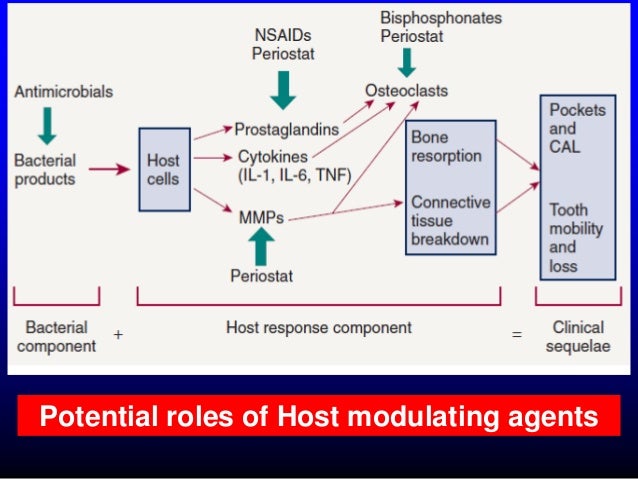
MMPI, matrixmetalloproteinaasi-inhibiittori Periostat


https://image.slidesharecdn.com/antiinfective-hostmodulationdralaa-140501041459-phpapp02/95/antiinfective-host-modulation-dr-alaa-34-638.jpg?cb=1398918053

MMPs , sinkistä riippuvia endopeptidaaseja

Sinkistä riippuvaiset endopeptidaasit MMPs

Anal Bioanal Chem. 2017 Aug 12. doi: 10.1007/s00216-017-0537-x. [Epub ahead of print]

Multimodal laser ablation/desorption imaging analysis of Zn and MMP-11 in breast tissues.

Abstract
Matrix metalloproteinases (MMPs) are a family of zinc-dependent endopeptidases. The main functions of these metalloproteinases are the degradation of the stromal connective tissue and basement membrane components.
Recent data from model systems suggest that MMPs are involved in breast cancer (BC) initiation, invasion, and metastasis.
Particularly, MMP-11 (stromelysin-3) is expressed in stromal fibroblasts adjacent to epithelial tumor cells, and high levels of this metalloproteinase were associated with tumor progression and poor prognosis of BC. Consequently, MMP-11 involved in these processes can be a candidate as a new potential prognostic biomarker in BC.
 Bioimaging techniques based on laser ablation/desorption and mass spectrometry are rapidly growing in biology and medicine for studies of biological systems to provide information of biomolecules (such as proteins, metabolites, and lipids) and metals with lateral resolution at the micrometer scale.
In this study, matrix-assisted laser desorption/ionization mass spectrometry imaging (MALDI-MSI) has been used for the first time to investigate the distribution of MMP-11 in human breast cancer tissues in order to show a possible correlation between cancerous and healthy samples, by differential proteomics and using such differences for possible cancer diagnosis and/or prognosis.
Additionally, those human breast tissue samples were analyzed in parallel by laser ablation inductively coupled plasma mass spectrometry (LA-ICP-MS) in order to gather additional information about the elemental distribution of Zn and its possible associations with MMPs.

KEYWORDS:

Biological samples; Laser ablation; Mass spectrometry/ICP-MS
PMID:
28801789
DOI:
10.1007/s00216-017-0537-x