Etiketter

fredag 15 mars 2019

ADAMTS- proteinaasit alaryhmineen. . Miten ADAMTS proteinaasit eroavat MMP ja ADAM- proteinaaseista rakenteellisesti?

https://arthritis-research.biomedcentral.com/articles/10.1186/ar1783
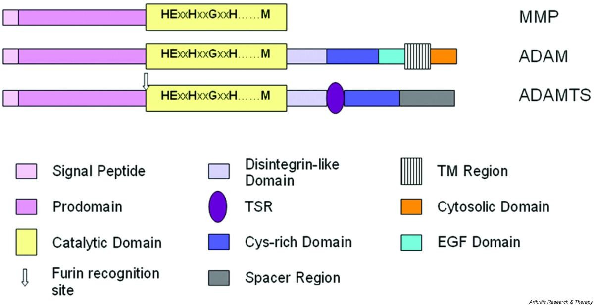
 Minimaalisinta kaavamaista domaanien ärjestäytymsitä kuvataan alla. Siinä on MMP, ADAM ja ADAMTS  esitettynä domaaneineen.
MMP, metalloproteinaasi
ADAM, eräs  disintegriini. ja metalloproteinaasi
ADAMTS,  eräs disintegriini ja metalloproteinaasi, jossa on trombospondiinidomeeni (TSR).
Huomioi, että useimmilla MMP-proteinaaseilla on lisäksi C-terminaalisia pidentymiä, joissa on  hemopexiinin-kaltaisia domaneja ja fibronektiini II-tyyppisiä domeeneja.
ADAMTS proteinaasit omaavat  0 - 14 kpl   trombospondiini tyyppi 1:n kaltaisten motiivien toistoja  (TSR) C-terminaali   Spacer  alueeseen. 

 Figure 1 Schematic representation of the minimal domain organisation of matrix metalloproteinase (MMP), ADAM (a disintegrin and metalloproteinase) and ADAMTS (a disintegrin and metalloproteinase with thrombospondin motifs; for example ADAMTS-4) proteinases. Note that most MMPs possess additional C-terminal extensions containing domains such as hemopexin-like and fibronectin type II domains. ADAMTS possess from 0 to 14 additional thrombospondin type 1-like repeat (TSR)-like motifs   C-terminal to the spacer domain.
 EGF, epidermal growth factor;
TM, transmembrane.
https://media.springernature.com/full/springer-static/image/art%3A10.1186%2Far1783/MediaObjects/13075_2005_Article_1662_Fig1_HTML.jpg

 https://arthritis-research.biomedcentral.com/articles/10.1186/ar1783

 ADAMTS- perheen jäsenet (  disintegriini ja metalloproteinaasi, jolla on trombospondiinimotiiveita) unnetaan siitä, että se vaikuttaa kehitykseen, angiogeneesiin, koagulaatioon ja artriitin progredioitumsieen.
 Proteinaasina niillä on seuraavia substraatteja:
  • von Willebrand-faktorin prekursori, edeltäjäproteiini,
  • ECM- komponentit( extrasellularisen matriksin komponentit)  kuten prokollageeni, hyalektaanit ( hyaluronaania sitovat proteoglykaanit, aggrekaani niiden joukossa)
  • dekoriini
  • fibromoduliini
  • ruston oligomeerinen matriksiproteiini


 ADAMTS-proteinaasien pitoisuudet  ja aktiviteetit  omaavat monitasoisen säätelyjärjestelmän  ja ne säätyvät   geeni-ilmentymän kontrollista, mRNA-pleissauksesta,  proteiinin prosessoitumisesta  ja TIMP- metalloproteinaasien kudosestäjien vaikutuksesta . Ihmisen rustoseulontatutkimukset ovat osoittaneet, että  moninaiset ADAMTS-perheen jäsenet saattavat  olla tärkeitä sidekudoksen homeostaasissa ja patologiassa.

(Suomennosta tiivistelmästä) 

  • Abstract

  • Members of the ADAMTS (a disintegrin and metalloproteinase with thrombospondin motifs) family are known to influence development, angiogenesis, coagulation and progression of arthritis. As proteinases their substrates include the von Willebrand factor precursor and extracellular matrix components such as procollagen, hyalectans (hyaluronan-binding proteoglycans including aggrecan), decorin, fibromodulin and cartilage oligomeric matrix protein. ADAMTS levels and activities are regulated at multiple levels through the control of gene expression, mRNA splicing, protein processing and inhibition by TIMP (tissue inhibitor of metalloproteinases). A recent screen of human cartilage has shown that multiple members of the ADAMTS family may be important in connective tissue homeostasis and pathology.

 ADAMTS-proteinaasien evoluutiosta ja rakenteesta.

  • ADAMTS evolution and structure

 Vuonna  1997 kuvasi Kuno kollegoineen ensimmäisen kerran hiiriltä ADAMTS-proteinaaseja. Sen jälkeen niitä on havaittu  eri nisäkkäiltä ja myös C-elegans-lajista.  Ne muodostavat osan  adamalysiineistä,  erään adamalysiinien  alaperheen B, perheen M12,  metallopeptidaasien  MA- heimossa ( määriteltynä MEROPSin tietueen mukaan). Rakenteellisesti ja evolutionaalisesti ne ovat  ADAM- proteinaasien kaltaisia (  disintegriini- ja metallopeptidaasidomaanit omaavia) . Myös ADAM- perhe on adamalysiinialaperheen entsyymejä.  Sukulaisuus on  etäisempää  MMP- proteinaaseihin (matriksimetalloproteinaaseihin (MMP, perhe M10 , heimo MA). Ylläoleva kuva näyttää  näiden kolmen alaperheen domaanirakenteita.

  • ADAMTS proteinases were first described in mice by Kuno and colleagues in 1997 [4] and have subsequently been identified in mammals and Caenorhabditis elegans. They form part of subfamily B (adamalysin subfamily), family M12, in clan MA of the metallopeptidases, as defined in the MEROPS database [5, 6] and are structurally and evolutionarily related to the ADAM (a disintegrin and metalloproteinase; also part of the adamalysin subfamily) enzymes and, more distantly, the matrix metalloproteinase (MMP; family M10 in clan MA) enzymes. A comparison of the minimal characteristic domain organisation of these groups of proteinases is shown in Fig. 1.

ADAMTS-geenien koodaamat proteiinit

 Ihmiseltä on tunnsitettu  19 eri ADAMTS- geenituotetta. Fylogeneettisiä ja geenianalyysiä  on tehty näistä sekvenssien perusteella ja ADAMTS- proteiinit voidaan jakaa yleisesti ottaen  neljään alaryhmään, joissa havaitaan myös rakenteellisia ja aktiviteetille ominaisia  piirteitä .  Tästä on myös kuva 2.  KAtalyyttisistä domeeneistä , sekvensseistä , tehty dendrogrammi  osoitaa miltei identtisiä domeeneja, mikä  osoitaa että katalyyttiset ja  niitä edistävät domeenit ovat evoloituneet  yhdessä .

1. Ensimmäinen  ADAMTS-ryhmä käsittää ADAMTS-1,-4,-5,-8,-9,-15, ja -20. ja se jakautuu kahteen  alaryhmäänjoista
 toisessa on  ADAMT-9 ja ADAMTS-20 ja
  muut mainitut ovat toisessa alaryhmässä ( ADAMTS-1,-4,-5,-8,-15).

2. Toinen selvästi erotettavissa oleva ADAMTS  ryhmä käsittää ADAMTS-2, -3 ja -14.

3,  Kolmannessa  ryhmässä on pelkästään ADAMTS-13.

4. Neljännessä,  hatarammin määritellyssä  ryhmässä ovat muut jäljellä olevat. ne ovat edelleen luokiteltu  neljään pariin rakenteellisten piirteiden perusteella:
ADAMTS-19 ja -17,
ADAMTS-18- ja 16,
ADAMTS-12 ja -7,
ja ADAMTS -10 ja -6.
On myös julkaistu  yksityiskohainen  tutkimus  ADAMTS-perheen jäsenten välisistä fylogeneettisistä suhteista.

  •  Nineteen distinct human ADAMTS gene products have been identified. A nearest-neighbour dendrogram constructed (using ClustalW 1.7 [7]) from sequence alignments of the entire protein indicates that human ADAMTS proteins can be broadly divided into four subdivisions, which also seem to share structural characteristics and activities (see Fig. 2 and below). A dendrogram constructed from the sequence alignment of the catalytic domains was almost identical, which implies that the catalytic and ancillary domains evolved together (data not shown). The first of the divisions, consisting of ADAMTS-1, -4, -5, -8, -9, -15 and -20, subdivides into two further groups, one composed of ADAMTS-9 and -20 and the other of ADAMTS-1, -4, -5, -8 and -15. A second, well-defined, subgroup contains ADAMTS-2, -3 and -14. ADAMTS-13 stands alone, and the remaining ADAMTS members form a loosely defined subgroup within which members are further divided into four pairs (ADAMTS-19 and -17, ADAMTS-18 and -16, ADAMTS-12 and -7, and ADAMTS-10 and -6) sharing structural features. A detailed study of the phylogenetic relationship of the ADAMTS family members has recently been published [8].

ADAMTS domain structure

The signal sequence of ADAMTS proteins is followed by a pro-region of varying length, but which is unusually short in ADAMTS-13. The pro-domain of all ADAMTS proteinases contains at least one furin cleavage consensus motif; it is therefore generally believed that the zymogen forms of ADAMTS proteinases are cleaved intracellularly and that secreted proteins are in the mature form. This mechanism of maturation is supported by studies of ADAMTS-4, which identify an N terminus of F213ASLS in supernatants conditioned by cells transfected with ADAMTS-4, suggesting that the prodomain is efficiently removed in vivo [9]. The same study also demonstrated that purified proADAMTS-4 could be cleaved by recombinant furin in cell-free experiments. Furin has recently been shown to interact with the pro-form of ADAMTS-4 and to co-localise within the trans-Golgi network [10]. Using furin inhibitors and RNA interference techniques, the removal of the pro-domain was inhibited without affecting secretion, demonstrating an important role for furin in intracellular processing [10]. The same study also revealed the presence of furin-independent pro-domain processing pathways in some cells.

 The catalytic domains of ADAMTS proteinases share a high degree of similarity and contain the zinc-binding sequence HEXXHXXGXXH, in which the catalytic zinc is coordinated by the three histidine residues. This arrangement is facilitated by the conserved glycine, which permits a tight hairpin loop and enables the third histidine to occupy its correct position [11, 12]. As in all MMPs and adamalysins, the zinc-binding sequence is followed at a short distance C-terminally by a conserved methionine residue, an active-site arrangement that has been termed 'metzincin-type'. This methionine constitutes the 'Metturn', a tight turn arranged as a right-handed screw that seems to serve an important function in the structure of the active site [11].

 The catalytic domain is followed by a region with 25 to 45% identity to the snake venom disintegrins, although it does not contain the cysteine arrangement of the latter [13]. This domain has therefore been termed disintegrin-like, though there is currently no published evidence that this ADAMTS domain interacts with integrins.


Unlike ADAM proteins, ADAMTS proteinases possess a well-conserved thrombospondin type 1-like repeat (TSR), homologous to the type I repeats of thrombospondins 1 and 2 [14], between the disintegrin-like and cysteine-rich domain (CRD). By analogy to thrombospondins 1 and 2 [15], the central TSR of ADAMTS proteinases is believed to function as a sulphated glycosaminoglycan-binding domain. The independently expressed central TSR of murine ADAMTS-1 required 0.46 to 0.66 M NaCl for elution from a heparin affinity column, indicating that this motif forms a functional heparin-binding unit [16].


Unlike ADAM proteins, ADAMTS proteinases possess a well-conserved thrombospondin type 1-like repeat (TSR), homologous to the type I repeats of thrombospondins 1 and 2 [14], between the disintegrin-like and cysteine-rich domain (CRD). By analogy to thrombospondins 1 and 2 [15], the central TSR of ADAMTS proteinases is believed to function as a sulphated glycosaminoglycan-binding domain. The independently expressed central TSR of murine ADAMTS-1 required 0.46 to 0.66 M NaCl for elution from a heparin affinity column, indicating that this motif forms a functional heparin-binding unit [16].

The CRD is a well-conserved cysteine-rich sequence containing 10 cysteine residues. In contrast to ADAM proteins, in which the CRD is followed by epidermal growth factor (EGF)-like repeats, a transmembrane domain and C-terminal cytosolic region, all ADAMTS proteinases possess instead a cysteine-free 'spacer' region. This domain varies in length and contains several conserved hydrophobic residues in the N-terminal portion and an extremely variable C-terminal portion. The expression of various domain-deletion constructs of murine ADAMTS-1 revealed the CRD-spacer sequence as a functional extracellular matrix (ECM)-binding domain [16]. This role was supported by investigation of C-terminally processed forms of human ADAMTS-4, which, in combination with a deletion construct lacking the CRD-spacer sequence, indicated that these domains also bind to both heparin and the glycosaminoglycans of aggrecan (predominantly keratan and chondroitin sulphates) [9]. Three putative heparin-binding sequences were identified within the CRD-spacer sequence of ADAMTS-4, one within the CRD and two within the spacer, and peptides corresponding to these sequences were shown to inhibit the binding of ADAMTS-4 to heparin [9].


With the exception of ADAMTS-4, which terminates after the spacer region, all ADAMTS proteinases possess between 1 and 14 TSRs C-terminal to the spacer region (Fig. 2). The sequence of these additional TSRs is more variable between the ADAMTS proteinases than is the central TSR, but the independent expression of the C-terminal TSRs of murine ADAMTS-1 has indicated that these motifs can form functional heparin-binding units [16]. The TSRs of the C-terminal region are arranged in one, two or three tandem arrays. Between arrays is either a short linker sequence (ADAMTS-9 and ADAMTS-20) or a mucin-like domain (ADAMTS-7 and ADAMTS-12) [17].


Four additional types of module have been described in the ADAMTS group and all are present C-terminal to the TSR arrays. ADAMTS-9 and -20 contain a unique module, also found in the C. elegans ADAMTS GON-1, containing 10 conserved cysteine residues [18]. Several ADAMTS proteinases (-6, -7, -10, -12, -16, -17, -18 and -19) possess a PLAC (protease and lacunin) domain containing six conserved cysteine residues, which is found in some pro-protein convertases [19]. A C-terminal extension containing a unique embedded PLAC domain is present in ADAMTS-2, -3 and -14. Finally, CUB (complement C1r/C1s, Uegf (EGF-related sea urchin protein) and BMP-1 (bone morphogenic protein-1)) domains are present at the C terminus of ADAMTS-13 [20]. This domain is also present in spermadhesins, tumour necrosis factor-stimulated gene-6 and the complement proteins C1r, C1s and mannan-binding lectin-associated serine proteinases, among others [21], and there is evidence to suggest that these domains mediate protein-protein interactions with other CUB domain-containing proteins [22, 23].

torsdag 14 mars 2019

MMP, matriksimetalloporteinaasi, TIMP, kudosvälitilan proteaasi-inhibiittori

Duodecim lehti julaksi  kymmenen vuota siten artikkelin eräästä MMP-matriksiproteinaasista MMP.8  ja sen kudosvälitilaestäjästä TIMP-1.

https://www.duodecimlehti.fi/lehti/2007/22/duo96896

Seerumin kollagenaasi 2:n (MMP-8) ja kudosvälitilan estäjä 1:n (TIMP-1) pitoisuudet ateroskleroosin riskitekijänä miehillä

2007;123(22):2673
Arterioscler Thromb Vasc Biol


Serum matrix metalloproteinase-8 concentrations are associated with cardiovascular outcome in men
Anita M. Tuomainen1, Kristiina Nyyssönen2, Jari A. Laukkanen2, Taina Tervahartiala1, Tomi-Pekka Tuomainen2, Jukka T. Salonen3, Timo Sorsa1, Pirkko J. Pussinen1
Arteriosclerosis, Thrombosis, and Vascular Biology 2007; Oct 11; [Epub ahead of print]

Elimistön krooniset tulehdukset, kuten Chlamydia pneumoniae -infektio ja parodontiitti, lisäävät riskiä sairastua sydän- ja verisuonitauteihin (SVT). Tulehduksien aikana matriksin metalloproteinaasit (MMP:t) osallistuvat soluväliaineen ja tyvikalvojen muokkaamiseen. Lisäksi ne ilmentyvät ateroskleroottisissa leesioissa, missä ne heikentävät leesiota suojaavaa kollageenirakennetta. MMP-8 on ensimmäisiä kollageenien hajottamisessa ilmentyviä metalloproteinaaseja. Sen toimintaa estää proteaasi-inhibiittori TIMP. Seerumin MMP-8- ja TIMP-1-pitoisuuksien vaikutuksesta sydän- ja verisuonitautien ilmaantumiseen ei ole tehty pitkäaikaisia seurantatutkimuksia.
Tämän tutkimuksen hypoteesit olivat, että seerumin MMP-8- ja TIMP-1-pitoisuudet liittyvät sydän- ja verisuonisairauksiin ja että suurentuneet pitoisuudet ennustavat SVT-tapahtumien riskiä. Potilasaineistona oli Kuopio Ischaemic Heart Disease Risk Factor (KIHD) -aineisto. Se koostui 1 018:sta 50-68-vuotiaasta miehestä, jotka vastasivat vuosina 1991-93 elämäntapoihin liittyviin kysymyksiin ja joilta mitattiin verenpaine, painoindeksi ja kaulavaltimon seinämän paksuus (IMT) sekä kerättiin seeruminäytteet. Kymmenen vuoden seuranta-aikana rekisteröitiin tutkimuksen päätetapahtumat: akuutti sydäninfarkti tai kuolema.
Tulosten mukaan seerumin suurentunut MMP-8- tai MMP-8/TIMP-1-pitoisuus oli suurempi miehillä, joilla oli tutkimuksen alussa SVT tai subkliininen ateroskleroosi (IMT vähintään 1 mm). Miehillä, joilla ei ollut SVT:tä tutkimuksen alussa, MMP-8-pitoisuuden kasvu 20 µg/l (1 SD) lisäsi molempien päätetapahtumien riskiä noin 15 %. Miehillä, joilla oli tutkimuksen alussa subkliininen ateroskleroosi, suuri MMP-8-pitoisuus ennusti SVT-kuolemaa riskisuhteella 3,03 (kuva).
Tutkimuksen perusteella seerumin MMP-8-pitoisuus saattaa olla kehittyvän ateroskleroosin ja sepelvaltimotaudin merkittävä biomarkkeri sekä ennustaa huonoa lopputulosta sydän- ja verisuonitaudeissa.
1Helsingin yliopiston hammaslääketieteen laitos ja HUS:n suu- ja leukasairauksien klinikka; 2Kuopion yliopiston kansanterveystieteen ja kliinisen ravitsemustieteen laitos; 3Oy Jurilab Ltd

Arvioi artikkeli

(* ) (* ) ( *) (* ) (* )

MMP/MT-MMP nimistä, substraateista ja inhibiittoreista. Treenaus ja obsesitas aspekti!


https://www.dovepress.com/cr_data/article_fulltext/s103000/103877/img/VHRM_103877_T001.jpg

 Abstract: Matrix metalloproteinases (MMPs) are zinc- and calcium-dependent endoproteinases that have the ability to break down extracellular matrix. The large range of MMPs’ functions widens their spectrum of potential role as activators or inhibitors in tissue remodeling, cardiovascular diseases, and obesity.
 In particular, MMP-1, -2, and -9 may be associated with exercise and obesity. Thus, the current study reviewed the effects of different types of exercise (resistance and aerobic) on MMP-1, -2, and -9. Previous studies report that the response of MMP-2 and -9 to resistance exercise is dependent upon the length of exercise training, since long-term resistance exercise training increased both MMP-2 and -9, whereas acute bout of resistance exercise decreased these MMPs. Aerobic exercise produces an inconsistent result on MMPs, although some studies showed a decrease in MMP-1. Obesity is related to a relatively lower level of MMP-9, indicating that an exercise-induced increase in MMP-9 may positively influence obesity. A comprehensive understanding of the relationship between exercise, obesity, and MMPs does not exist yet. Future studies examining the acute and chronic responses of these MMPs using different subject models may provide a better understanding of the molecular mechanisms that are associated with exercise, obesity, and cardiovascular disease.

Keywords: cardiovascular disease, gelatinases, collagenases, TIMP


I nhibition of MMPs

Two types of MMP inhibitors exist: endogenous and exogenous inhibitors.

 Tissue inhibitors of metalloproteinase (TIMPs) are endogenous inhibitors that can be secreted (TIMP-1, TIMP-2, TIMP-4) or bound to ECM components (TIMP-3).20 They inactivate MMPs by forming bonds with catalytic zinc in 1:1 ratios within the MMP structure.21 


 They do so by creating noncovalent interactions between the N-terminal domain of the TIMP and the active site of MMPs. Recent studies have shown TIMPs’ therapeutic potential in cardiovascular diseases (CVDs)

The MMP-inhibitory effects of TIMP-1 involve binding of C-terminal domain to pro-MMP-2 and pro-MMP-9. TIMP-1 may specifically help ECM manipulation in ischemia in such a way that it may be a surrogate marker for increased ECM turnover.22

TIMP-2 reverses ECM remodeling in human cardiac tissue in a dose-dependent manner.23

 TIMP-3 prevented degradation of cardiac tissue matrix after a myocardial infarction by reducing MMPs in vascular smooth muscle cells.24

 Changes in the ECM of atrial fibroblast in rheumatic heart disease have been associated with TIMP-4 expression.25
Exogenous inhibitors include hydroxamic acid derivative and thiirane gelatinase inhibitor SB-3CT on several MMPs.2629 Batimastat (BB-94), a hydroxamic acid derivative, has been recently found to treat aneurysms on using nanoparticle technology to directly administer anti-MMP target to abdominal aorta aneurysm.30 In rat aorta cell culture, administration of BB-94 decreased 90% of MMP-9 activity and 10% of MMP-2 activity, while showing no effect on TIMP-2 activity.
The effect of change in MMP activity was tested by injecting BB-94 directly into the abdominal aorta. The blank control showed a 269% increase in the aneurysm, while the treatment group showed only a 40% increase in expansion.
Another derivative, mastmariat, has shown success as a therapeutic drug in repressing non-small-cell lung cancer (NSLC) in a Phase I trial.31 Oral administration of marimastat was added to the accepted treatment with carboplatin and paclitaxel in order to test whether or not marimastat would affect the kinetics of the treatment. TIMPs and hydroxamic acid derivatives play significant roles in general physiology and pathology, and thus can develop as a therapeutic target in the future.

"Major sheddases" ADAM-10 ja ADAM-17.. Mikäa on sheddaasi? Wikipedia artikkelin suomennsota

Wikipedia: SHEDDASE
https://en.wikipedia.org/wiki/Sheddase

SHEDDAASIT  ovat kalvoon sitoutuneita entsyymejä, jotka pystyvät aiirä sijainnistaan  pilkkomaan irti kalvon läpi ulottuvien muiden  proteiinien solunulkoista osaa, ektodomeenia, liukoiseksi kappaleeksi. KATSO KUVA:
Nämä sheddaasientsyymit voivat täten aktivoida kalvon läpi ulottuvia proteiineja, jos kyse on reseptoriproteiineista ( esim HER2) tai ne voivat  leikata irti osan  kalvon läpäisevästä proteiinista  silloinkin kun se on jo sitoutuneena agonistiinsa ja niin katkaistu palanen voi  vaikuttavan agonistin kanssa  ( esim EGFR) päästä stimuloimaan jonkin toisen solun reseptoria.
Pääsheddaaseja ovat ADAM-10 ja ADAM-17 ja  niitä  vastaan kehitetyt sheddaasi-inhibiittorit   voivat  vahvistaa syövänvastaista terapiaa.

Sheddases are membrane-bound enzymes that cleave extracellular portions of transmembrane proteins, releasing the soluble ectodomains from the cell surface. Many sheddases are members of the ADAM or aspartic protease (BACE) protein families.[1]
These enzymes can activate a transmembrane protein if it is a receptor (e.g., HER2), or cut off the part of the transmembrane protein which has already bound an agonist (e.g., in the case of EGFR), allowing this agonist to go and stimulate a receptor on another cell.  Sheddase inhibitors active on ADAM10 and ADAM17 can potentiate anti-cancer therapy.[2] .


 https://en.wikipedia.org/wiki/Sheddase#/media/File:Ectodomain_shedding_en.svg

Sheddaasin funktiosta  (Functions)

 On oletettu, että sheddaasien aktiivisuutta tapahtuu  suhteessa yleiseen  entsymaattisen aktiivisuuteen. Tutkimukset  viittaavat siihen, että sen sijaan  sheddaasit  olisivat  suhteessa  fosfatidyyliseriinialtistukseen.  ( Fosfatidyyliseriini, PS,  solun ulkopinnalla fosfolipidikalvossa on  solun ikääntymisen merkki) . Artikkelissa kerrotaan eräästä tutkimuksesta PSA-3 soluilla:  Kun näiden solujen  kyky syntetisoida fosfatidyyliseriiniä aleni, havaittiin sheddaasiaktiviteetin vähentyneen. kun solut jälleen pystyivät syntetisoimaan fosfatidyyliseriiniä, sheddaasiaktiivisuus palasi. Tästä tutkijat tekivät johtopäätöksen, että fosfatidyyliseiinialtistus olisi välttämätön soluille, jota ne voivat ilmentää sheddaasiaktiivisuutta.
 ( Oma kommentti: tätä PS-näkökohtaa en ole ennen  huomannut. Solun fosfolipidien  homeostaasissa on flip-flop-scrambling mekanismi, jossa  kaikki kalvolipidit   siirtyvät omiin suhteellisiin asemiinsa ja tätä tasapainoa ylläpitää  kalvon energiatila ja kalvopotentiaali, koko  ihmiskehossa riippuu tästä normaalsita kalvoorganisaatiosta.  Fosfatidyyliseriinin pitäisi normaalisti sijaita  kalvon sisäosiin päin ja vanhenevassa kalvossa se nousee pintaan, mikä  antaa merkin siitä,  että solu on valmis fysiologiseen apoptoitumiseen. Saattaa olla että tämä aktivoi disintegraaseja nopeasti  avustamaan  ja hajoittamaan  solukerroksia monitahoisesti - niillä on kymmenittäin substraatteja -  ja altistamaan solun  tulehduksettomaan katoamiseen, jota fysiologinen apoptoosi on. Epäfysiologiseen apotoosiin liittyy tulehdustekijä moni patologia).

  • It has been postulated that the activity of sheddases occurs in relation to the amount of general enzymatic activity. Research indicates that sheddases are instead related to phosphatidylserine exposure. When PSA-3 cells' ability to synthesize phospatidylserine was repressed, sheddase activity decreased, and the sheddase activity returned to normal levels when the cells were again able to synthesize phosphatidylserine. This led researchers to conclude that phosphatidyserine exposure is necessary for cells to exhibit sheddase activity.[3]

Voiko sheddaaseja hyödyntää kliinisesti?  (Uses)

 Sheddaasientsyymien  mekanismien ja funktioiden takia niitä on  tutkittu tarkoituksella löytää mahdollisia lääketieteellisiä käyttöjä. yksi tällainen käyttömahdollisuus  olisi allergisten vasteiden ja immuunijärjestelmän prosessien hoito.
ADAM10 vastaa  muun muassa  CD23-immunoglobuliinireseptorin  ektodomaanin irrottamisesta  ja tästä seuraa liukoista  sCD23, jota esiintyy sitten seerumissa ja se taas  vaikuttaa immuunivasteita. Joillain yksilöillä voi alkaa tulehduksellinen tauti kuten astma. Ottaen huomioon , että ADAM10 sheddaasientsyyminä pilkkoo irti liukoista  (soluble)  CD23:a  ja lisää liukoisen , kiertävän sCD23:n pitoisuuksia,  on mahdollista hoitaa  aiheutuvia tauteja kohdistetusti  estämällä sheddaasifunktiota.

  • Due to the nature of the mechanisms and functions of sheddase enzymes, they have been studied on the basis of discovering possible uses in medicine. One such use is in the treatment of allergic responses and other processes of the immune system. ADAM10 is responsible for the shedding of the CD23 Immunoglobulin receptor, which releases soluble sCD23.[4] sCD23 present in the blood serum contributes to immune response and, to some, the onset of inflammatory disease such as asthma. Given that ADAM10 sheddase cleaves CD23 and increases the levels of sCD23, possible treatments for these diseases may center around the inhibition of sheddase function. 
  ADAM17 eli TACE, tuumorinekroosifaktori alfaa konvertoiva entsyymi  on sellainen sheddaasientsyymi,. jota on havaittu monen tyypin syövissä ja se  voisentakai toimia biologisena merkitsijänä syövän havaitsemisvaiheessa. TACE- ilmentyminen ei  suoranaisesti korreloi  mihinkään syöväna steeseen, muta kuitenkin on havaittu että  tään sheddaasientsyymin aktiivisuus on merkitsevästi  esiintyvää pään ja kaulan alueen syövissä normaaleihin  viljeltyihin soluihin verrattuna.
  • Tumor necrosis factor alpha converting enzyme (TACE) is a sheddase protein that has been observed in many types of cancer and could serve as an important Biomarker (medicine) used in the detection of cancer.[5] While the expression of TACE does not directly correlate with particular stages of cancer, the shedding activity of the enzyme is significantly more prominent in head and neck cancer cells compared to normal cultured cells.[5]
 .Kommentti Wikipedia-artikkeli  antaa alaviitteen  jossa esiintyy lisätietoa  näistaä ADAM10 ja ADAM17- -sheddaaseista. otan siitä sitaatin tähän  allaolevaan otsikkoon.  SHADDASE- Artikkeli on kirjoitettu 2017 aikaan ja mainittu että se on STUB.  Tämän jälkeen on tieto  kaikista ADAM-proteiineista  kasvanut tieto geeneistä tarkentunut PubMed. Muutamista muistakin ADAM- proteaaseista  mainitaan sheddaasiominaisuutta.  Mutta ADAM10 ja ADAM17 erottuvat rakenteen yksityiskohdan (disintegriinidomeenin) puolesta  muista.

  1. See also

    It has become clear in the past years that ectodomain shedding is an initial step for the activation of specific receptors such as Notch, ErbB-4 and the angiopoietin receptor Tie-1. Notch-1 signaling is essential for endothelial differentiation, and tumor angiogenesis, while the angiopoietin receptor Tie-1 facilitates embryonic blood vessel formation.[12][13] Upon binding of their ligands, Notch-1 and Tie-1 undergo proteolytic cleavage of the ectodomains by ADAM17 and ADAM10. This cleavage frees the cytoplasmic fragment for cellular signaling, in the case of Notch-1, it transfers to the nucleus. Many cytokines and growth factors are synthesized as membrane bound proforms which undergo proteolytic shedding for activation. The ephrins EPH receptor A2 and A3 are shed by ADAM10 creating cleaved soluble Eph receptors, which inhibit tumor angiogenesis in mice.[14] Additional examples are the proteolytic shedding of soluble E-selectin,[15] shedding of urokinase receptor (uPAR) by MMP-12 creating soluble uPAR which has chemotactic properties for leukocytes and progenitor cells, and the shedding of interleukin-6 receptors by ADAM10 and ADAM17 which facilitates interleukin-6 signaling in endothelial cells.[16] Semaphorin 4D is cleaved from its membrane bound form by MT1-MMP (MMP-14) in tumor cells; it then interacts with plexin B1 on endothelial cells promoting pro-angiogenic chemotaxis.[17] Shedding of a membrane anchored cytokine or growth factor by ADAM proteinases may be relevant for various signal transduction events. Alternatively, shedding may be required for the ligand to diffuse to distant receptors. Shedding may be required for the down regulation of signals by removing signaling ligands, or cleavage and release of receptors. Release of the receptor may also generate soluble receptors which act as decoys by sequestering ligands. These findings indicate that ectodomain shedding is a ubiquitous process facilitating a wide variety of cellular events involved in angiogenesis. Because potent biological modifiers are generated, it is likely controlled by highly regulated mechanism. Along with ADAMs and MT-MMPs, membrane bound serine proteases also may play a role in ectodomain shedding.

    References

  2. R&D Systems (Winter 2006). "Need help at the cell surface? Ask your local sheddase". Cytokine Bulletin.
    Sheddases cleave membrane proteins at the cell surface, releasing soluble ectodomains with altered location and function. Some sheddases are membrane proteins themselves that belong to metalloprotease (ADAM and MMP) or aspartic protease (BACE) families. Their activity can be constitutive or regulated through various processes such as PKC activation, Ca2+ influx, and lipid rafts.1-3
    A single sheddase may cleave a variety of substrates. A classic example in this category, ADAM17, was initially identified as TNF-alpha-converting Enzyme (TACE), and is known to shed a variety of growth factors, receptors, and adhesion molecules.1,4 This suggests that overall conformations of the substrates are more important than primary amino acid sequences in determining the accessibility for cleavage by sheddases.
    Multiple sheddases can cleave the same substrate. Under certain circumstances this may result in different consequences. ADAM17, ADAM10, and MMP-14/MT1-MMP are all known to shed CD44, an adhesion molecule that interacts with hyaluronic acid in the ECM.2 Additionally, amyloid precursor protein (APP) processing by alpha, or by beta- and gamma-secretases has differential effects on the production of alpha beta peptide, a major plaque component found in brains of Alzheimer’s disease patients. Cleavage of APP by beta-secretase (BACE-1 and -2) creates a substrate for gamma-secretase, resulting in alpha beta peptide production. In contrast, alpha-secretase (ADAM10, 17 and 9) cleavage between the beta- and gamma-secretase sites prevents alpha beta peptide production.5 Juxtamembrane cleavage that creates a substrate for further processing that results in the release of a cytoplasmic domain has been termed regulated intramembrane proteolysis (RIP). Notch processing by ADAM10 RIP liberates the Notch intracellular domain, allowing it to translocate to the nucleus and activate the transcription of target genes.6
  3. Healthvalue: Sheddases and ADAMs
  4. Sommer, Anselm; Kordowski, Felix; Büch, Joscha; Maretzky, Thorsten; Evers, Astrid; Andrä, Jörg; Düsterhöft, Stefan; Michalek, Matthias; Lorenzen, Inken (2016-05-10). "Phosphatidylserine exposure is required for ADAM17 sheddase function". Nature Communications. 7. doi:10.1038/ncomms11523. ISSN 2041-1723. PMC 4866515. PMID 27161080.
  5. "ADAM10 is a principal 'sheddase' of the low-affinity immunoglobulin E receptor CD23" (PDF). Retrieved November 5, 2016.
  6.  
  7. "Sheddase Activity of Tumor Necrosis Factor- Converting Enzyme Is Increased and Prognostically Valuable in Head and Neck Cancer". Retrieved 2016-11-05.
 

Kehitetty ADAM17-inhibiittori ZLDI-8, joka inhiboi Notchsignaloinnin HCC soluissa(2018)

http://www.probechem.com/products_ZLDI-8.aspx
Tämä ei ole   kliininen lääke.
 Zhang Y et al. 2018:

ZLDI-8 (ZLDI8;IAC-8)

Catalog No.: PC-35373Not For Human Use, Lab Use Only.
ZLDI-8 (IAC-8) is a novel Notch signaling pathway inhibitor for Notch activating/cleaving enzyme ADAM-17, significantly decreases the level of NICD and accumulation of NICD in the nucleus.

 ZLDI-8 (IAC-8) is a novel Notch signaling pathway inhibitor for Notch activating/cleaving enzyme ADAM-17, significantly decreases the level of NICD and accumulation of NICD in the nucleus; exhibits cytotoxic activity against MHCC97-H cells with IC50 of 5.32 uM, reduces the expression of pro-survival/anti-apoptosis regulators, Survivin and cIAP1/2, also increases the expression of epithelial marker E-Cadherin and reduces mesenchymal markers N-Cadherin and Vimentin in HCC cells; significantly disrupted the activity of Notch pathway in HCC cells and inhibits the epithelial-mesenchymal transition (EMT) process of HCC cells; ZLDI-8 treatment enhances the susceptibility of HCC cells to Sorafenib, Etoposide, and Paclitaxel both in vitro and in vivo.

 Related  products. Target Notch
http://www.probechem.com/target_Notch.aspx

Tässä lähteessä mainitaan muita Notch- inhibiittoreita, mutta niistä mikään ei ole varsinaisia ADAM17-inhibiittoreita.

Muistiin  14.3. 2019

onsdag 13 mars 2019

ADAM- molekyyleistä

https://www.researchgate.net/publication/301638469_A_Disintegrin_and_Metalloprotease_ADAM_Historical_Overview_of_Their_Functions

  • Nives Giebeler

    Suomennosta tiivistelmästä 

     Senjälkeen kun  oli löydetty  ensimmäinen disintegriiniproteiini käärmeen myrkystä ja oli myös tunnistettu nisäkkäiltä kalvoon ankkuroitunut metalloproteinaasi-disintegriini, jolla on tehtäviä fertilisaatiossa, seurasi kolme vuosikymmentä tutkimusten aikaa ja tunnistettiin  runsaasti näiden  proteinaasiperheiden jäseniä ja useita biokemiallisia mekanismeja, jotka säätivät niiden ilmenemistä solussa  ja aktiivisuutta. 
    Mikä tärkeintä  on  myös tunnistettu näiden proteiinien  in vivo-funktioita- kuten  niiden osallistuminen solujen jakaantumiseen kehityksen aikana,  niiden tulehdsreaktioita moduloiva vaikutus  ja niiden osallistuminen syövän syntyyn. 
    Tässä katsauksessa  tehdään yleisnäkymää ADAM-proteaasien perheestä  ( ADAM on lyhennys  englantilaisista sanoista , jotka tarkoittavat:  "eräs disintegriini ja metalloproteaasi") ja valaistaan  muutamia  tärkeimpiä tutkimussaavutuksia ADAM-proteinaasien  analyyseistä , mitkä painottavat näiden proteiinien  tärkeyttä  fysiologisissa ja patologisissa prosesseissa  ajan mittaan. 
    • Abstract
    Since the discovery of the first disintegrin protein from snake venom and the following identification of a mammalian membrane-anchored metalloprotease-disintegrin implicated in fertilization, almost three decades of studies have identified additional members of these families and several biochemical mechanisms regulating their expression and activity in the cell. Most importantly, new in vivo functions have been recognized for these proteins including cell partitioning during development, modulation of inflammatory reactions, and development of cancers. In this review, we will overview the a disintegrin and metalloprotease (ADAM) family of proteases highlighting some of the major research achievements in the analysis of ADAMs' function that have underscored the importance of these proteins in physiological and pathological processes over the years.
    Muistiin  13.3. 2019  

    KLOMEMNTTI:
    Näitä ADAM-proteiineja on luomakunnassa   monilla, mutta ihmisillä  on lueteltu  numeroon 33 asti, vaikka  myöhemmin on jouduttu tekemään pieniä muutoksia  luetteloon, tarkennettu, jos onkin kyse pseudogeenistä  ja yhdistetty jokin  ADAM -proteiini saman  nimen alle. moni ADAmgeeni esiintyy  klustereina. . 

    PubMed hakulaiteesta Geenien  luettelosta löytyy seuraavia ihmisten ADAM- proteinaaseja. 
    Ihmisen ADAM-proteiinit ja geenit tänään 13.3. 2019 PubMed hakuna ja  yhtenvetona kromosomittain. 

    1. Kromosomi

    ADAM15,  (1q21.3)  MDC15
     https://www.ncbi.nlm.nih.gov/protein/NP_001248393.1
    vain tällä metargidiini ADAM- metalloproteinaasilla on RGD- aminohapposekvenssi disintegriinidomeenissa ja sen takia  se  voi sitoa integriiniä.
    ADAM30,  (1p12) ,svph4
     https://www.ncbi.nlm.nih.gov/gene/11085
    Tämä geeni  on  testisspesifinen. 

    2.  Kromosomi

    ADAM17, (2p25.1)  CD156B, CSVP, NISBD, NISBD1, TACE  (TNFalpha converting enzyme, TNFa Convertase) .
     https://www.ncbi.nlm.nih.gov/pubmed/30069943
    Kehitetty inhibiittori ZLDI-8, joka estää ADAM17- ja  samalla Notch-signalointia, koska Notch on eräs  ADAM17- substraateista. 

    ADAM23, (2q33.3)  MDC3
    https://www.ncbi.nlm.nih.gov/gene/8745
    Geeni ilmenee aivoissa ja sydämessä eniten. . 

    4 Kromosomi

    ADAM29, ( 4q34.1) CT73 (cancer/testis antigen 73) , svph1
    https://www.ncbi.nlm.nih.gov/gene/11086 
    ADAM 29 ja ADAM7 ovat usein mutatoituneet melanomassa. 

    5 Kromosomi

    ADAM19. (5q33.3)  FKSG34, MADDAM (Metalloproteinase And Disintegrin Dendritic Antigen Marker) , MLTNB (Meltrin beta), Dendriittisolun kypsymisen merkitsijä.
    https://www.ncbi.nlm.nih.gov/gene/8728

    7 Kromosomi

    ADAM22, (7q21.12)   EIEE61, MDC2 (Early Infantile Epileptic Encephalopathy 61)
    Tämä ADAM22 ei omaa sinkkiä sitovaa motiivia ja sen takia siltä puuttuu  metalloproteinaasiominaisuus. Sitä esiintyy paljon aivosissa ja arvellaan että  se toimii integriiniligandina. Hiirillä sen on havaittu  vaikuttavan virheetöntä myelinisaatiota ja tarkkaa  synaptista  transmissiota.   Funktion  disruptio aivoissa vikuuttaa AMPA-reseptorien  funktiota , mikä selittää osaltaan   epileptisyyttä.
    https://www.ncbi.nlm.nih.gov/gene/53616

    8. Kromosomi

    ADAM2 ( 8p11.22), CRYN1, CRYN2,CT15 (cancer/testis antigen 15), FTNB ( fertilin subunit beta), , PH-30-P, PH30, PH-30-beta
     https://www.ncbi.nlm.nih.gov/gene/2515

    ADAM3A, pseudogeeni   (8p11.22) , ADAM3, CYRN1 (cyritestin 1) , IMDC1, restricted expr. towards testis(pseudogene). Homozyg. puute: pons gliomariski.
    https://www.ncbi.nlm.nih.gov/gene/1587

    ADAM5, (8p11.22) ADAMSP, TMDCII, restricted towards testis, pseudogene.
    * Cluster:  8p21.2  :  ADAM7, ADAM28, ADAMDEC1:

    *ADAM7 (8p21.2) , ADAM-7, EAP1 (epididymal apical protein 1) , GP-83, GP83(sperm maturation related glycoprotein 83)  Mutatoitunut   ADAM7 usein  melanomassa.
     https://www.ncbi.nlm.nih.gov/gene/8756

    ADAM- perheen alaperheeseen kuuluu    immuunifunktion hankkinut decysiini,
    Klusterin  jäsenillä on  samankaltaisia omnaisuuksia,.
    • *ADAMDEC1 (8p21.2)  (decysin ) https://www.ncbi.nlm.nih.gov/pubmed/12037602/ 
    •  https://www.ncbi.nlm.nih.gov/gene/10863  
    • Metzincin- metalloproteinaasi.  Mieto proteinaasi.  Tuottaa  aktiivia EGF  verihiutaleista. tärkeä monosyyttien erilaistumisessa  ja DC solujen solujen kypsymisessä.  Vaikuttaa  reologiaan. Kiinnostava linkki! 2000 löytynyt alaperhe. Se eroaa ADAM-perheestä siten, että  siltä puuttuu osittain disintegriinidomeeni ja  cysteiinidomeeni puuttuu kokonaan, mistä nimi: decysiini. Tämä geeni on  evoloitunut  geeniduplikaatiolla  metalloproteinaasigeeniklusterissa  8p12  kohalla.  Vaikka  tämä geeni menetti metalloproteinaasiominaisuuksia,  se hankki immuunifunktioita.

     *ADAM28  (8p21.2) MDC-L (metalloproteinaasin kaltainen, disintegriinin kaltainen- cysteiinipitoisen kaltainen proteiini) MDCL, MDCII, eMDCII (epididymal) . Omaa immuunifunktiota ja kuuluu yllämainittuun geeniklusteriin. 
    https://www.ncbi.nlm.nih.gov/gene/10863
    ADAM9 (8p11.22)  CORD9 (Cone rod dystrophy 9) , MCMP  (Myeloma Cell metalloproteinase) , MDC9, Mltng
    https://www.ncbi.nlm.nih.gov/gene/8754
    ADAM18, (8p11.22)  ADAM27, tMDCIII ( Mature sperm surface protein)
    https://www.ncbi.nlm.nih.gov/gene/8749

    ADAM24 (8p22), replaced with gene ADAM24P, pseudogeeni
    https://www.ncbi.nlm.nih.gov/gene/8745

    ADAM32 (8p11.22) , MMP-12- kaltainen, testiskudosproteiini Li13.
    https://www.ncbi.nlm.nih.gov/gene/203102

    10. Kromosomi

    ADAM8 (10q26.3) , CD156, CD156a, MS2 (solupinta-antigeeni) , ihmisen leukosyyttidifferentiaation antigeeni,  osallistuu temozolomide- kemoresistenssiin.
    https://www.ncbi.nlm.nih.gov/gene/101

    ADAM12, (10q26.2)  ADAM12-OT1, CAR10, MCMP, MCMPMltna, MLTN, MLTNA (meltrin alpha)(Maternal serum marker for pre-natal development)
    https://www.ncbi.nlm.nih.gov/gene/8038 

    12. Kromosomi

    ADAM1A, pseudogeeni  /12q24.12q24.13.   ADAM1, ADAM1P, FTNAP, Ftna, PH-30a, Fertilin alpha, pseudogene https://www.ncbi.nlm.nih.gov/gene/8759

    14. Kromosomi

    ADAM6, pseudogeeni(14q32.33) , tMDCIV, C14orf96
    https://www.ncbi.nlm.nih.gov/gene/?term=ADAM6
     
    ADAM20 (14q24.2) 
     fertiliini alfan  kaltainen, testisspesifinen. kuten ADAM21
     https://www.ncbi.nlm.nih.gov/gene/8748
     ADAM21 (14q24.2)
    fertiliini alfan kaltainen, myöstestisspesifinen. Myös nimi: ADAM31.

    15- Kromosomi

    ADAM10, (15q21.3) , AD10, AD18, CD156c, CDw156, HST-187, MADM (mammalian disintegrin metalloprioteinase), RAK (reticulate acropigmentation of  Kitamura) , Kuz ( Kuzbanim protein homolog)
    Tämä metalloproteinaasi  proteiini pilkkoo TNF-alfaa ja E-cadherinia.
     https://www.ncbi.nlm.nih.gov/gene/102

    17. Kromosomi

    ADAM 11,  (17q21.31). MDC ( Metalloproteinase, Disintegrin, Cystein- rich protein). Proteiinia ilmenee runsaasti  runsaasti  aivoissa. Geenin  sijainnin takia rintasyövän kandidaattigeenejä.

    20. Kromosomi

    ADAM33 (20p13), C20orf153, DS964F7.1
    https://www.ncbi.nlm.nih.gov/gene/80332
    Tämä proteiiniassosioituu astmaan ja hyperreaktiivisuuteen. 

    Muistiin  14.3. 2019 


ADAM10 , alfa-sekretaasi , monitoiminen metalloproteaasi aivojen alueella

https://www.ncbi.nlm.nih.gov/pubmed/265229
2015 Dec;135:1-20. doi: 10.1016/j.pneurobio.2015.10.003. Epub 2015 Oct 29.

The alpha secretase ADAM10: A metalloprotease with multiple functions in the brain.

(Suomennosta 13.3. 2019)  Tiivistelmä  (Abstract)

 Disintegriinien ja  metalloproteinaasien perheeseen kuuluva ADAM10 on kalvoon ankkuroitunut proteiini, joka pystyy pilkkomaan useita kalvoon sitoutuneita proteiineja prosessissa, jota sanotaan ektodomaanin irrottamiseksi, ectodomain shedding. 

 Keskushermostossa ADAM10 on herättänyt suurinta mielenkiintoa, kun se kuvattiin amyloidin prekursoriproteiinin (APP)  alfa-sekretaasina  yli 10 vuotta sitten. Innostuttiin pitämään ADAM10-proteiinia mahdollisena uutena lääkekohteena AD- taudissa, vaikka ADAM10-proteiinin fysiologiset funktiotkin olivat vielä vähemmän  käsitettyjä. Hiirillä ADAM10-puute on  alkiovaiheessa letaali ja tämä johtuu siitä, että  ADAM10 -funktiota tarvitaan Notch reseptoriproteiinien  pilkkomiseen ja signalointiin.  Notch on toinen ADAM10:n susbtraatti.
 
Uudempien  ehdollisesti ADAM10-vajeisten hiirien kehittämisen avulla  on saatu lisätietoa tästä proteiinista.  On tunnistettu uusia ADAM10-substraatteja aivoista,  ja niin on paljastunut hämmästyttävän lukuisia ja fundamentaalisia funktioita ADAM10- proteiinille sekä alkion aivojen kehityksessä että aikuisen  neuronaalisen verkoston homeostaasissa.
 
Mekanistisesti  ADAM10 kontrolloi näitä funktioita  käyttämällä hyödyksi  keskushermoston ainutlaatuisia postsynaptisia substraatteja, erityisesti synaptisia soluadheesiomolekyylejä kuten neuroligiini-1, N-cadheriini, NCAM, Efriini-A2 ja Efriini-A5.
 
 Johdonmukaisesti  ADAM10-aktiviteetin  säätelyvika liittyy psykiatrisiin ja neurologisiin tauteihin kuten epilepsiaan,   fragile X- oireyhtymään ja Huntingtonin tautiin. Tässä katsauksessa valaistaan nykyistä edistymistä keskushermoston ADAM10:n  substraattien ja  funktioiden , säätelyn ja solubiologian  ymmärtämisessä . Pohditaan myös sen merkitystä aivoperäisten tautien lääkekohteena. 

  • Proteins belonging to the 'A Disintegrin And Metalloproteinase' (ADAM) family are membrane-anchored proteases that are able to cleave the extracellular domains of several membrane-bound proteins in a process known as 'ectodomain shedding'. In the central nervous system, ADAM10 has attracted the most attention, since it was described as the amyloid precursor protein α-secretase over ten years ago. Despite the excitement over the potential of ADAM10 as a novel drug target in Alzheimer disease, the physiological functions of ADAM10 in the brain are not yet well understood. This is largely because of the embryonic lethality of ADAM10-deficient mice, which results from the loss of cleavage and signaling of the Notch receptor, another ADAM10 substrate.
  •  However, the recent generation of conditional ADAM10-deficient mice and the identification of further ADAM10 substrates in the brain has revealed surprisingly numerous and fundamental functions of ADAM10 in the development of the embryonic brain and also in the homeostasis of adult neuronal networks. 
  • Mechanistically, ADAM10 controls these functions by utilizing unique postsynaptic substrates in the central nervous system, in particular synaptic cell adhesion molecules, such as neuroligin-1, N-cadherin, NCAM, Ephrin A2 and A5. Consequently, a dysregulation of ADAM10 activity is linked to psychiatric and neurological diseases, such as epilepsy, fragile X syndrome and Huntington disease. This review highlights the recent progress in understanding the substrates and function as well as the regulation and cell biology of ADAM10 in the central nervous system and discusses the value of ADAM10 as a drug target in brain diseases.
  • Copyright © 2015 Elsevier Ltd. All rights reserved.

KEYWORDS:

ADAM;
 APP;
Alzheimer disease;
Axon;
Cell adhesion;
Ectodomain shedding;
 Neurite outgrowth;
 Neuronal differentiation;
 Notch-1;
Post synapse;
Prion;
Protease;
 Signaling;
 Synaptogenesis;
Therapy
PMID:
26522965
DOI:
10.1016/j.pneurobio.2015.10.003
[Indexed for MEDLINE]