Etiketter

fredag 23 mars 2018

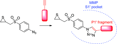
Gelatinaasi-inhibiittorien kehittelystä MMP-2 ja MMP-9-inhibitio. Thiirane

http://pubs.rsc.org/en/content/articlelanding/2014/ra/c3ra46402d#!divAbstract

New clicked thiirane derivatives as gelatinase inhibitors: the relevance of the P1′ segment

Abstract

Gelatinases (MMP-2 and MMP-9), a subfamily of Matrix Metalloproteinases (MMPs), are involved in several pathologies and especially in cancer. Thiirane is a latent-zinc binding group used for the design of potent inhibitors of gelatinases. Here we report a new family of thiirane inhibitors, obtained by click chemistry. Thus, an azide fragment containing the thiirane group was connected to several lipophilic alkynes, which were designed to interact with the S1′ pocket of the two gelatinases. Our hit compound (2f) displayed submicromolar inhibition of MMP-2 (IC50 = 0.62 μM). Computational studies have been used to compare the binding mode of compound 2f in MMP-2 with the reference thiirane inhibitor (SB-3CT), allowing us to discuss the relevance of the P1′ segment in order to maximize potency.
Graphical abstract: New clicked thiirane derivatives as gelatinase inhibitors: the relevance of the P1′ segment

Supplementary files

Publication details

The article was received on 04 Nov 2013, accepted on 01 Apr 2014 and first published on 03 Apr 2014

Article type: Paper
DOI: 10.1039/C3RA46402D
Citation: RSC Adv., 2014,4, 17726-17735


torsdag 22 mars 2018

Anti-proteaasi-proteaasi tasapaino hengitysteissä

Esimerkkinä kystinen fibroosi:

https://www.researchgate.net/figure/The-importance-of-the-balance-between-proteases-and-antiproteases-in-CF-airways-The_fig1_44579199

The main source of protease activity in the CF lung is thought to be activated neutrophils, however, the role of proteases derived from other cell sources, such as mononuclear and epithelial cells, as well as exogenously derived bacterial proteases may also play a vital role in mediating destruction of the lung as seen in CF (as illustrated in Fig. 1). The main emphasis of this review will be to address the role of these endogenous and exogenous proteases and describe past, present and future developments in the search for an efficient antiprotease therapy in CF.

https://www.researchgate.net/profile/Cliff_Taggart/publication/44579199/figure/fig1/AS:203132643614727@1425442004177/The-importance-of-the-balance-between-proteases-and-antiproteases-in-CF-airways-The.png


The release of proteolytic enzymes during the inflammatory response has enormous potential to exacerbate and prolong inflammation causing numerous deleterious effects such as lung tissue destruction and reduced bacterial clearance.

To counteract this, the airways are equipped with a highly regulated antiprotease shield to dampen and control excessive proteolytic activity. The protease-antiprotease imbalance hypothesis of chronic neutrophil-mediated lung disease contends that this imbalance, or overwhelming of the natural antiprotease defence mechanisms, determines pulmonary phenotype [31].

 Several CF studies have correlated protease lung burden with disease severity and have shown an inverse relationship to the status of the antiprotease defence shield [7, 32-34].

The function of the airway antiprotease defence system is to inhibit the activity of cognate proteases thereby preventing potentially damaging degradation of host tissue (Fig. 1).

The primary antiproteases of the airway are AAT, secretory leucoprotease inhibitor (SLPI), elafin, TIMPs and cystatins [31, 35-38]. The primary protease targets of AAT, SLPI and elafin are the serine proteases, NE, proteinase 3 and cathepsin G, all of which are released by activated neutrophils [30, 31, 35, 36].

MMP-12- inhibittoori

Muistiin:
http://www.merckmillipore.com/SE/en/product/MMP-12-Inhibitor-MMP408-CAS-1258003-93-8-Calbiochem,EMD_BIO-444291?ReferrerURL=https%3A%2F%2Fwww.google.se%2F

Synonyms: (S)-2-(8-(Methoxycarbonylamino)dibenzo[b,d]furan-3-sulfonamido)-3-methylbutanoic acid

Date of issue:
16.11.2014
Version:
1.0
Company
EMD Millipore Corporation | 290 Concord Road, Billerica, MA 018
21, United
States of America | General Inquiries: +1-978-715-4321 | Monday
to Friday, 9:00
AM to 4:00 PM Eastern Time (GMT-5)
Millipore S.A.S. (Merck Millipore European Headquarters) | 39 R
oute Industrielle
de la Hardt, Molsheim 67120, Fr
ance | General Inquiries: +39 (0
) 33 90 46 90 00
| Monday to Friday, 8:00 AM to 4:00 PM Central European Time (G
MT+1)
Emergency telephone
number
800-424-9300 CHEMTREC (USA)
+1-703-527-3887 CHEMTREC (International)
24 Hours/day; 7 Days/week
Catalogue No.:
444291
Product name:
MMP-12 Inhibitor, MMP408
REACH Registration
Number:
A registration number is not available for this substance as the substance
or its use are exempted from registration according to Article2 REACH
Regulation (EC) No 1907/2006, the annual tonnage does not require a
registration or the registration is envisaged for a later registration deadline.
This item is not a hazardous substance and does not contain hazardous ingredients, substances with European Community workplace exposure limits or substances of v
ery high concern (SVHC) above their respective disclosure limits. Hence a safety data sheet is not required according to Regulation(EC) No. 1907/2006 (REACH) and also not available in this case....

onsdag 21 mars 2018

MMP klusterin aktivaatiossa plasmiini merkitsevä.

Plasminergisen systeemin tehtävänä on  normalisoida kehoa sen jälkeen, kun on ilmentynyt fibrinogeenistä fibriiniä, jota pitäisi hajoittaa. Samalla plasmiini  useassa tapauksessa  joutuu aktivoimaan avuksi MMP-klusterin, jotta  suuresta hyytymästä päästään eroon  ja kudoksia voidaan uudistaa.

Mistä syystä siten  kehon koagulaatiojärjestelmä  aktivoituu ja trombiini alkaa pilkkoa  fibrinogeenistä fibriiniä  ja vaikutaa  loputla  palautumatonta  stabiilia fibriiniä?
 Siihen on tiestysti paljon syitäja  niihin on  suurin osa  tieteellisestä artikkeleista reologian alalta keskittynytkin.

Tromboosin estämiseen tavataan käyttää milloin  aspiriinia, milloin  marevan tyyppistä antikoagulanttia , milloin muita   strategioita , kuten trombosyyttien estäjiä.
Mutta keholla on myös oma järjestelmänsä, jolla se aivan jatkuvasti hoitaa  negatiivisen antikoagulaation   ja järjestelmän pitäisi  normaalissa kehossa toimia.  Siinä trombiinin ilmenemä  aktivoi veressä normaalisti  kiertävää  C-proteiinia ja  aktivoitu C-proteiini aPC toimii  koagulaatiokaskadin rauhoittajana.

 Mutta jos käytetään  suuria määriä aspiriinia  tai marevania,  C-proteiinia ei saada aktivoitumaan antikoagulantiksi, joten joudutaan  pilleri pillerin jälkeen säätämään  lopulta koko järjestelmää  sen sijaan että luottaisiin kehon normaaliin  koagulatiiviseen tasapainoon.

Mitä se normaali tasapaino käsittää?  Eräs tärkeä koagulaatiotekijä on aktiivi kalsiumjoni. (Tekijä FIV)
 Jota kalsiumjoni pysyisi  normaalipuitteissa ja kertyisi  varastoihinsa kuten luustoon,   todellakin tarvitaan tehokasta liikuntaa.
Jos liikunta puuttuu, on miltei mahdoton  ilman lääkkeitä selvitä koagulaatiotasapainon hoidosta ja samalla vielä luusto purkaa kalsiumia.
Jos haluaa  säilyttää normaalin koagulaatio-järjestelmänsä kannattaa kohtalaisen tai runsaan  liikunnan lisäksi  käyttää  K1-vitamiinipitoista ravintoa, kuten vihreitä vihanneksia ja kasvisöljyjä, jolloin  saa edellytyksiä  negatiivisen kogulaatiokontrollin jäsenten posttranslationaaliseen  muokkautumiseen funktionaaliseen muotoon.  Tämä voisi  "pinnan alla" näkymättömissä  pitää  reologista tasapainoa sellaisessa tilassa, että  ei muodostuisi  siinä määrin  sulamatonta fibriiniä, että plasminerginen järjestelmä aktivoituu ja  jopa aktivoi MMP-järjestelmän.

 Normaalisti MMP-järjestelmä on  hyvin  vähäisesti aktivoituneena.  Vain tarvittessa jossain paikallisesti se vaikuttaa  uudistelemassa solukalvojen extrasellulaarista materiaalia- se hajoittaa monin spesifisin tekijin vanhat materiaalit, jota uutta pääsee muodostumaan.

Jotta riehahtanutta  MMP-järjestelmää saa rauhoittumaan  täytyy katsoa syntyjä syviä aivan fibriinin  muodostumiseen asti.  Tämä on hyvin  yksinkertaistettu selitys.

 MMP-järjestelmän rauhoittamisessa ei ole eduksi käyttää  antitromboottisia lääkkeitä kuten ASA ja  antikoagulanttia vaikka lyhyellä tähätimellä ne  estävät fibriiniä muodostaumasta.
MMP  on hyvin monimutkainen järjestelmä eikä sille ole olemassa vielä mitään erityisiä  rauhoittavia lääkkeitä, estäjiä. ne eivät vähene  ASA tai antikoagulanttilääkityksestä.

Nykyään vielä kartoitetaan, miten paljon järjestelmä riehuu erilaisissa tiloissa.

Sellaisen liikunnan , joka ei ledeeraa, riko  kudoksia  (aiheuta kudosrikkoja),  ei varinaisesti pitäisi  aktivoida MMP-järjestelmää ylimäärin.

Tässä  lähinnä korostaisin  kudoksille aivan  perustavasti tärkeitä  olosuhteita:  ravintosuositukset  ovat sitä linjaa, kasvisten osuutta vuosi vuodelta korostetaan enemmän ja se samalla stabiloi  Kvitamiinin saannin hieman korkeammalle tasolle, mitä ennen näitä suosituksia.  Suosituksissa on myös sellaisia öljyjä, jotka antavat K1-vitamiinia.  Tämä on hidas tie  korjata  villiintynyttä MMP-klusteritoimintaa ja toivottavasti  tapahtuu jotain palautumista - siis  MMP-järjestelmän  palautumista  inaktiiviin tilaan.

Tämä on uusi kartta joka on ilmeentynyt koagulaatiokaskadin ja fibrinolyyttisen kaskadin  vierelle, eikä  kaikki vuorovaikutukset ole millään tavalla  varmoja  nuolia kartalla vielä. Lisätekijä on TIMP-kartta, MMP-kudosinhibiittorit , jotka näyttävät jo  riehuvan  syöpätutkimusalan karttojen puolella. nekin ovat yllättäen osoitatneet pahat puolensa, eikä nekään ole  mitenkään helposti lääkkeellisesti säädeltäviä.

Alun perin saataa koko tämä ihmeellisen  hyvä kaskadisekvessi mennä pieleen vain pelkästä liikunnan puutteesta ja joistain  elintapatekijöistä.  Pelkkä  puolen  tunnin lisäliikunta päivää kohti voisi olla  avain  oikeaan suuntaan johtavalle ovelle.


Matrixmetalloproteinaasit keuhkofibroosissa (IPF)


https://www.ncbi.nlm.nih.gov/pubmed/26121236

Am J Respir Cell Mol Biol. 2015 Nov;53(5):585-600. doi: 10.1165/rcmb.2015-0020TR.

Matrix metalloproteinases as therapeutic targets for idiopathic pulmonary fibrosis.

Craig VJ1,2, Zhang L1, Hagood JS3,4, Owen CA1,5

Abstract

Idiopathic pulmonary fibrosis (IPF) is a restrictive lung disease that is associated with high morbidity and mortality. Current medical therapies are not fully effective at limiting mortality in patients with IPF, and new therapies are urgently needed. Matrix metalloproteinases (MMPs) are proteinases that, together, can degrade all components of the extracellular matrix and numerous nonmatrix proteins. MMPs and their inhibitors, tissue inhibitors of MMPs (TIMPs), have been implicated in the pathogenesis of IPF based upon the results of clinical studies reporting elevated levels of MMPs (including MMP-1, MMP-7, MMP-8, and MMP-9) in IPF blood and/or lung samples. Surprisingly, studies of gene-targeted mice in murine models of pulmonary fibrosis (PF) have demonstrated that most MMPs promote (rather than inhibit) the development of PF and have identified diverse mechanisms involved. These mechanisms include MMPs: (1) promoting epithelial-to-mesenchymal transition (MMP-3 and MMP-7); (2) increasing lung levels or activity of profibrotic mediators or reducing lung levels of antifibrotic mediators (MMP-3, MMP-7, and MMP-8); (3) promoting abnormal epithelial cell migration and other aberrant repair processes (MMP-3 and MMP-9); (4) inducing the switching of lung macrophage phenotypes from M1 to M2 types (MMP-10 and MMP-28); and (5) promoting fibrocyte migration (MMP-8). Two MMPs, MMP-13 and MMP-19, have antifibrotic activities in murine models of PF, and two MMPs, MMP-1 and MMP-10, have the potential to limit fibrotic responses to injury. Herein, we review what is known about the contributions of MMPs and TIMPs to the pathogenesis of IPF and discuss their potential as therapeutic targets for IPF.

KEYWORDS:

fibrosis; idiopathic pulmonary fibrosis; interstitial lung disease; lung; matrix metalloproteinase
PMID:
26121236
PMCID:
PMC4742954
DOI:
10.1165/rcmb.2015-0020TR
[Indexed for MEDLINE]
Free PMC Article

Clinical Relevance

In this Translational Review, we describe the molecular and cell biology of matrix metalloproteinases (MMPs) and tissue inhibitors of metalloproteinases, and review the evidence that links MMPs to idiopathic pulmonary fibrosis (IPF), the cellular sources of MMPs, and the mechanisms involved. Although initial studies of randomized clinical trials for nonselective MMP inhibitors as new therapies for cancer produced disappointing results, since then, newer approaches to target metalloproteinases more selectively have been developed for other diseases. We have included a discussion of the advantages (and potential limitations) of these new therapeutic approaches targeting MMPs and their potential as therapeutics for IPF.

Idiopathic Pulmonary Fibrosis

In the United States, approximately 50,000 patients are newly diagnosed with idiopathic pulmonary fibrosis (IPF) each year. The median survival of patients with IPF is only 3–5 years (1). Although numerous medical therapies have been evaluated in patients with IPF, the only therapies that slow the progression of this disease, pirfenidone (2) and nintedanib (3), are associated with side effects and are not fully effective at reducing mortality. Thus, there is an urgent need to identify novel therapeutic targets for IPF. Herein, we review the evidence linking members of the matrix metalloproteinase (MMP) family to the pathogenesis of IPF, identify knowledge gaps in the field of MMPs and IPF, and discuss potential approaches to target MMPs as novel therapeutics for IPF.
IPF is characterized by the deposition of excessive amounts of extracellular matrix (ECM) proteins in the lungs, thereby replacing the normal architecture of the lung. IPF is the most common type of idiopathic interstitial pneumonia, and is characterized pathologically by the pattern of usual interstitial pneumonitis. Although the etiology of IPF is still unclear, several pathogenic mechanisms have been implicated in its development, including aberrant repair of injured epithelium, fibroblast activation, epithelial-to-mesenchymal transition (EMT), collagen deposition, and immune cell dysfunction. MMPs are expressed by most of the cellular culprits and pathologic processes implicated in IPF pathogenesis.

MMPs

MMPs are zinc-dependent endopeptidases that, together, degrade all components of the ECM. Consequently, it was initially thought that MMPs would limit lung fibrosis by degrading ECM proteins in the lung. However, recent studies have implicated MMPs in regulating the activities of proteins other than ECM proteins, including mediators of inflammation, latent growth factors, antifibrotic growth factors, and cleaving cell surface molecules and receptors. However, most studies of MMP-deficient mice in pulmonary fibrosis (PF) models have shown the opposite—that MMPs promote pulmonary fibrotic responses to injury.

MMP Structure

MMPs are multidomain proteins (Figure 1). The signal peptide at the amino terminus targets the protein to the cell’s secretory pathway. The propeptide domain, containing the highly conserved cysteine switch motif, PRCGXPD, is cleaved during activation of the latent proenzyme by yet-to-be identified peptidases. The catalytic domain contains the highly conserved Zn2+-binding motif, HEXXHXXGXXH, in which the three histidines (H) bind to the active site zinc, and the nucleophilic glutamate (E) attacks the substrate’s peptide bond. The proline-rich hinge domain connects the catalytic domain to the C-terminal domain with a flexible segment of up to 75 residues. The carboxyterminal hemopexin-like domain regulates substrate binding and specificity. Some MMPs contain additional domans
.... kts. jatko  linkistä!

Mikä toimisi MMP klusterin inhibiittorina?

Biol Res Nurs. 2010 Apr;11(4):336-44. doi: 10.1177/1099800409346333. Epub 2009 Dec 22.

The role of doxycycline as a matrix metalloproteinase inhibitor for the treatment of chronic wounds.

Abstract

Many chronic wounds fail to heal with conventional therapy, resulting in disability and impaired quality of life. New technologies using recombinant growth factors, autologous growth factors, or bioengineered skin-tissue substitutes have been shown to be effective, but these treatments are costly. An effective, low-cost treatment to improve healing of chronic wounds is needed. The molecular environment of chronic wounds, like many other chronic inflammatory diseases, contains abnormally high levels of proinflammatory cytokines (tumor necrosis factor [TNF]-alpha and interleukin [IL]-1beta]) and matrix metalloproteinases (MMPs), which impair normal wound healing. In animal models and clinical studies of ulcerative diseases, doxycycline, an inexpensive and Food and Drug Administration (FDA)-approved antibiotic, appears to inhibit members of the MMP superfamily like MMPs and TNF-alpha-converting enzyme (TACE). This article provides an overview of the roles of MMPs and intrinsic tissue inhibitors of metalloproteinases (TIMPs) in wound healing and the damaging effects of chronically elevated levels of MMPSs in chronic wounds. It also explores the use of topical doxycycline, a synthetic MMP inhibitor (MMPI), to enhance healing of chronic wounds.

Proteaasi-antiproteaasi-epätasapaino keuhkofibroosissa

https://www.ncbi.nlm.nih.gov/pubmed/29518524

Matrix Biol. 2018 Mar 5. pii: S0945-053X(17)30471-7. doi: 10.1016/j.matbio.2018.03.001. [Epub ahead of print]

The impaired proteases and anti-proteases balance in Idiopathic Pulmonary Fibrosis.

Idiopathic Pulmonary Fibrosis (IPF) is a devastating chronic, progressive and irreversible disease that remains refractory to current therapies.

Matrix metalloproteinases (MMPs) and their inhibitors, tissue inhibitors of MMPs (TIMPs), have been implicated in the development of pulmonary fibrosis since decades. Coagulation signalling deregulation, which influences several key inflammatory and fibro-proliferative responses, is also essential in IPF pathogenesis, and a growing body of evidence indicates that Protease-Activated Receptors (PARs) inhibition in IPF may be promising for future evaluation.

Therefore, proteases and anti-proteases aroused great biomedical interest over the past years, owing to the identification of their potential roles in lung fibrosis. During these last decades, numerous other proteases and anti-proteases have been studied in lung fibrosis, such as matriptase, Human airway trypsin-like protease (HAT), Hepatocyte growth factor activator (HGFA)/HGFA activator inhibitor (HAI) system, Plasminogen activator inhibitor (PAI)-1, Protease nexine (PN)-1, cathepsins, calpains, and cystatin C.

 Herein, we provide a general overview of the proteases and anti-proteases unbalance during lung fibrogenesis and explore potential therapeutics for IPF.

KEYWORDS:

Anti-protease; Cysteine protease; Idiopathic Pulmonary Fibrosis; Matrix metalloproteinase; Serine protease
PMID:
29518524
DOI:
10.1016/j.matbio.2018.03.001

Outline

  1. Highlights
  2. Abstract
  3. Keywords
  4. Idiopathic Pulmonary Fibrosis (IPF)
  5. Pro- and anti-fibrotic lung proteases and anti-proteases deregulation in IPF
    • Matrix metalloproteinases (MMPs) and their cognate inhibitors
      • MMPs
      • TIMPs
    • Cysteine proteases and their cognate inhibitors
      • Cysteine proteases cathepsins, ATG4B, and calpains
      • Cystatin C
    • Serine proteases and their cognate inhibitors
      • Coagulation serine proteases
      • Type II transmembrane serine proteases (TTSPs)
      • The HGFA-HAI imbalance in IPF
      • Serine proteases inhibitors: plasminogen activator inhibitor-1 (PAI-1) and protease nexin-1 (PN-1) serpins
  6. Therapeutic targeting of proteases/anti-proteases for IPF
  7. Future directions
  8. Conflicts of interest
  9. Funding sources
  10. References

Keuhkofibroosin taustatekijöitä fibriinin homeostaasijärjestelmästä

https://www.ncbi.nlm.nih.gov/pubmed/29474926
Int J Biochem Cell Biol. 2018 Feb 21;97:108-117. doi: 10.1016/j.biocel.2018.02.016. [Epub ahead of print]

The fibrogenic actions of the coagulant and plasminogen activation systems in pulmonary fibrosis.

Abstract

Fibrosis causes irreversible damage to lung structure and function in restrictive lung diseases such as idiopathic pulmonary fibrosis (IPF). Extravascular coagulation involving fibrin formation in the intra-alveolar compartment is postulated to have a pivotal role in the development of pulmonary fibrosis, serving as a provisional matrix for migrating fibroblasts. Furthermore, proteases of the coagulation and plasminogen activation (plasminergic) systems that form and breakdown fibrin respectively directly contribute to pulmonary fibrosis. The coagulants, thrombin and factor Xa (FXa) evoke fibrogenic effects via cleavage of the N-terminus of protease-activated receptors (PARs). Whilst the formation and activity of plasmin, the principle plasminergic mediator is suppressed in the airspaces of patients with IPF, localized increases are likely to occur in the lung interstitium. Plasmin-evoked proteolytic activation of factor XII (FXII), matrix metalloproteases (MMPs) and latent, matrix-bound growth factors such as epidermal growth factor (EGF) indirectly implicate plasmin in pulmonary fibrosis. Another plasminergic protease, urokinase plasminogen activator (uPA) is associated with regions of fibrosis in the remodelled lung of IPF patients and elicits fibrogenic activity via binding its receptor (uPAR). Plasminogen activator inhibitor-1 (PAI-1) formed in the injured alveolar epithelium also contributes to pulmonary fibrosis in a manner that involves vitronectin binding. This review describes the mechanisms by which components of the two systems primarily involved in fibrin homeostasis contribute to interstitial fibrosis, with a particular focus on IPF. Selectively targeting the receptor-mediated mechanisms of coagulant and plasminergic proteases may limit pulmonary fibrosis, without the bleeding complications associated with conventional anti-coagulant and thrombolytic therapies.

KEYWORDS:

Coagulants; Factor X; Plasmin; Thrombin; Tissue factor; Urokinase plasminogen activator
PMID:
29474926
DOI:
10.1016/j.biocel.2018.02.016

torsdag 15 mars 2018

TGF reseptori ja Smad signalointi

https://www.youtube.com/watch?v=GuKjUearIUI
Tässä pienessä pätkässä näytetään perustava Smad signalointi ilman  estäviä  tekijöitä. TRIM33 voisi napata  ensinkin SMad4.n tuon yleis-Smadin ja monoubikitinyloida sen  niin ettei se voi tehdä kompleksia R-Smadien  kanssa ja päästä  tumaan.
TRIM33 voi myös napata  R-SMad2/3 ja mennä tumaan . Se kompleksi lukee kromatiinia ja   SMAD4 /Smad2/3 alkaa transkription.
TRIM33 on  corepressiivinen ja hidastaa tai modifioi  transkriptiota. 
Kts.
 SMAD geenit
SMAD-proteiiniperhe 
https://www.sciencedirect.com/topics/biochemistry-genetics-and-molecular-biology/smad

tisdag 13 mars 2018

TIMP-1 metabolisessa oireyhtymässä taustatekijöitä


https://www.ncbi.nlm.nih.gov/pubmed/16288749
FEBS Lett. 2005 Nov 21;579(28):6417-22. Epub 2005 Nov 2.

Proinflammatory adipocytokines induce TIMP-1 expression in 3T3-L1 adipocytes.

Abstract

Tissue inhibitor of metalloproteinase (TIMP)-1 is an adipocyte-secreted protein upregulated in obesity which promotes adipose tissue development. Furthermore, the proinflammatory adipocytokines tumor necrosis factor alpha (TNFalpha) and interleukin (IL)-6 induce insulin resistance, and plasma concentrations are increased during weight gain.
 In the current study, the impact of TNFalpha and IL-6 on TIMP-1 mRNA and protein expression was determined in 3T3-L1 adipocytes. Interestingly, TNFalpha and IL-6 induced TIMP-1 protein secretion more than 3- and 2-fold, respectively. Furthermore, TIMP-1 mRNA was upregulated in a time- and dose-dependent fashion. Inhibitor experiments suggested that nuclear factor kappaB and p 44/42 mitogen-activated protein kinase are involved in both, basal and adipocytokine-induced TIMP-1 expression. Moreover, the thiazolidinedione troglitazone partly reversed TNFalpha- but not IL-6-induced TIMP-1 synthesis. Taken together, we demonstrate that TIMP-1 expression is selectively upregulated in fat cells by proinflammatory adipocytokines and might play a role in maintaining adipose tissue mass in obesity.
PMID:
16288749
DOI:
10.1016/j.febslet.2005.10.033

Mitä muita funktioita TIMP-1 omaa?


http://www.biochemj.org/content/459/3/565.figures-only


Accelerated Publication

TIMP-1 modulates chemotaxis of human neural stem cells through CD63 and integrin signalling

Soo Youn Lee, Jung Mi Kim, Soo Young Cho, Hyun Suk Kim, Hee Sun Shin, Jeong Yong Jeon, Rukhsana Kausar, Seon Yong Jeong, Young Seek Lee, Myung Ae Lee

TIMP-1 geeni sijaitsee X-kromosomissa


https://www.ncbi.nlm.nih.gov/gene/7076
Aikanaan kun katselin  MMP-proteinaasien  aktivoitumisen kaskadia ja sitten sitä inhibitioreittiä TIMPs, siinä oli jollain tavalla puutteellista  karttakohtaa. Mikä  säätelee  negatiivisesti TIMP-inhibiittorit?

Näyttää olevan  paljon pahaa tästä TIMP-ylössäätymisestä kudoksissa:

TIMP-1 
Also known as
EPA; EPO; HCI; CLGI; TIMP; TIMP-1
Summary
This gene belongs to the TIMP gene family. The proteins encoded by this gene family are natural inhibitors of the matrix metalloproteinases (MMPs), a group of peptidases involved in degradation of the extracellular matrix. In addition to its inhibitory role against most of the known MMPs, the encoded protein is able to promote cell proliferation in a wide range of cell types, and may also have an anti-apoptotic function. Transcription of this gene is highly inducible in response to many cytokines and hormones. In addition, the expression from some but not all inactive X chromosomes suggests that this gene inactivation is polymorphic in human females. This gene is located within intron 6 of the synapsin I gene and is transcribed in the opposite direction. [provided by RefSeq, Jul 2008]
Expression
Broad expression in appendix (RPKM 344.6), gall bladder (RPKM 211.2) and 15 other tissues See more


TIMP-1 ja CD63 biomerkitsijöitä glioblastoomassa


https://www.ncbi.nlm.nih.gov/pubmed/29523123
TIMP-1 assosioituu kemoterapiaresistenssiin glioblastoomassa. 
BACKGROUND:
We have previously identified tissue inhibitor of metalloproteinases-1 (TIMP-1) as a prognostic marker in glioblastomas. TIMP-1 has been associated with chemotherapy resistance, and CD63, a known TIMP-1-binding protein, has been suggested to be responsible for this effect. The aim of this study was to assess CD63 expression in astrocytomas focusing on the prognostic potential of CD63 alone and in combination with TIMP-1.
METHODS:
CD63 expression was investigated immunohistochemically in a cohort of 111 astrocytomas and correlated to tumor grade and overall survival by semi-quantitative scoring. CD63 expression in tumor-associated microglia/macrophages was examined by double-immunofluorescence with ionized calcium-binding adapter molecule 1 (Iba1). The association between CD63 and TIMP-1 was investigated using previously obtained TIMP-1 data from our astrocytoma cohort. Cellular co-expression of TIMP-1 and CD63 as well as TIMP-1 and the tumor stem cell-related markers CD133 and Sox2 was investigated with immunofluorescence. TIMP-1 and CD63 protein interaction was detected by an oligonucleotide-based proximity ligation assay and verified using co-immunoprecipitation.
RESULTS:
The expression of CD63 was widely distributed in astrocytomas with a significantly increased level in glioblastomas. CD63 levels did not significantly correlate with patient survival at a protein level, and CD63 did not augment the prognostic significance of TIMP-1. Up to 38% of the CD63+ cells expressed Iba1; however, Iba1 did not appear to impact the prognostic value of CD63. A significant correlation was found between TIMP-1 and CD63, and the TIMP-1 and CD63 proteins were co-expressed at the cellular level and located in close molecular proximity, suggesting that TIMP-1 and CD63 could be co-players in glioblastomas. Some TIMP-1+ cells expressed CD133 and Sox2.
CONCLUSION:
The present study suggests that CD63 is highly expressed in glioblastomas and that TIMP-1 and CD63 interact. CD63 does not add to the prognostic value of TIMP-1. Co-expression of TIMP-1 an
d stem cell markers as well as the wide expression of CD63 might suggest a role for TIMP-1 and CD63 in glioblastoma stemness.

TIMP-4 matrixproteinaasiestäjä on prognostinen biomerkitsijä astrosytoomassa

Astrosytooman invaasioon liittyy eräät geenit:  TIMP-4 oli yliesiintyvä  pilosyyttisessä astrosytoomassa verrattuna diffuseihi astrosytoomiin.  O ajateltu että TIMP-4 olisi vaikutukseltaan tuumorin vastainen, muta tutkijat ovat tässä toista mieltä. TIMP-4 partneri on CD63.  Tutkijat analysoivat  471 gliomaa, joisasaoli 354 astrosytoomaa. Oligodendrogliomaan  verrattuna  astrosytoomassa olivat molemmat nämä merkitsijät koholla  samoin pilosyyttisessä astrosytoomassa verrattuna II asteen  diffuusiin astrosytoomaan.  TIMP-4 ja CD63  merkitsijöiden samanaikaisilmenemät astrosytoomassa osoitettiin tässä työssä ensimmäistä kertaa ja ne toimivat gliooman differentiaalidiagnostiikassa  astrosyyttisen fenotyypin  ilmentäjinä. Glioblastoomassa niiden esiintymällä on prognostinen merkitys.

https://www.ncbi.nlm.nih.gov/pubmed/20693981
Mod Pathol. 2010 Oct;23(10):1418-28. doi: 10.1038/modpathol.2010.136. Epub 2010 Aug 6.

TIMP-4 and CD63: new prognostic biomarkers in human astrocytomas.Rorive S1, Lopez XM, Maris C, Trepant AL, Sauvage S, Sadeghi N, Roland I, Decaestecker C, Salmon I.

Based on the molecular profiling of astrocytomas, we previously identified a series of genes involved in astrocytoma invasion. Of these, tissue inhibitor of metalloproteinase-4 (TIMP-4) was found to be overexpressed in pilocytic astrocytomas relative to diffuse astrocytomas of any histological grade. Although some data suggest that TIMP-4 may be an anti-tumoral actor in astrocytomas, recent findings challenge this concept. The present study aims to investigate the diagnostic and prognostic values of TIMP-4 and its putative partner CD63 in human astrocytomas. Tissue microarray and image analysis were first carried out to quantitatively analyze the immunohistochemical expression of these proteins in 471 gliomas including 354 astrocytomas. Pathological semi-quantitative scores of both markers' expression were then established and correlated to astrocytoma diagnosis and patient prognosis. TIMP-4 and CD63 expressions were both overexpressed in astrocytomas compared with oligodendrogliomas (P<0 .001="" a="" addition="" adverse="" and="" as="" associated="" association="" astrocytic="" astrocytoma="" astrocytomas="" cd63="" cells.="" co-expression="" compared="" conclusion="" contribution="" diffuse="" evidence="" factors="" first="" glioblastomas.="" glioblastomas="" gliomas.="" grade="" high="" highlights="" identified="" identifies="" ii="" in="" independent="" it="" markers="" of="" outcomes="" p="" patients="" phenotype="" pilocytic="" prognostic="" progression="" provides="" scores="" shorter="" survival.="" the="" this="" timp-4="" to="" tumor="" were="" with="" work="">

tisdag 6 mars 2018

Gliooman strategioita: "gain of function"

Astrogliaryhmällä Müllerin gliasoluilla retinassa on kykyä kestää oxidatiivista stressiä ja siinä järjestelmässä toimii glu-cystiini antiporter. jota  Glioomatuumorit  käyttävät saaden kykyä kestää oksidatiivista stressiä, kun  cystiinistä tehdään CSH:ta. Tässä alla on tutkittu  retinan Müllerin radiaalisten gliasolujen   osuutta antioksidanttisuudessa. Itse ajatelin että gliooman  kyky käyttää tätä glu css antiporteria  toi mieleen, että onko se lähtöisin näista retinan   alueen radiaalisista gliasoluista, jotka  tosiaan tarvitsevat korkean  antioksidanttisen kyvyn,  mitä itse asiassa  valolle ja UV.lle  altistumaton osa  aivon gliasolukkoa ei tarvitse.
Free Radic Biol Med. 2015 Sep;86:25-36. doi: 10.1016/j.freeradbiomed.2015.04.009. Epub 2015 Apr 25.

Sigma 1 receptor regulates the oxidative stress response in primary retinal Müller glial cells via NRF2 signaling and system xc(-), the Na(+)-independent glutamate-cystine exchanger.

Oxidative stress figures prominently in retinal diseases, including diabetic retinopathy, and glaucoma. Ligands for σ1R, a unique transmembrane protein localized to the endoplasmic reticulum, mitochondria, and nuclear and plasma membranes, have profound retinal neuroprotective properties in vitro and in vivo. Studies to determine the mechanism of σ1R-mediated retinal neuroprotection have focused mainly on neurons. Little is known about the effects of σ1R on Müller cell function, yet these radial glial cells are essential for homeostatic support of the retina. Here we investigated whether σ1R mediates the oxidative stress response of Müller cells using wild-type (WT) and σ1R-knockout (σ1RKO) mice. We observed increased endogenous reactive oxygen species (ROS) levels in σ1RKO Müller cells compared to WT, which was accompanied by decreased expression of Sod1, catalase, Nqo1, Hmox1, Gstm6, and Gpx1. The protein levels of SOD1, CAT, NQO1, and GPX1 were also significantly decreased. The genes encoding these antioxidants contain an antioxidant response element (ARE), which under stress is activated by NRF2, a transcription factor that typically resides in the cytoplasm bound by KEAP1. In the σ1RKO Müller cells Nrf2 expression was decreased significantly at the gene (and protein) level, whereas Keap1 gene (and protein) levels were markedly increased. NRF2-ARE binding affinity was decreased markedly in σ1RKO Müller cells. We investigated system xc(-), the cystine-glutamate exchanger important for synthesis of glutathione (GSH), and observed decreased function in σ1RKO Müller cells compared to WT as well as decreased GSH and GSH/GSSG ratios. This was accompanied by decreased gene and protein levels of xCT, the unique component of system xc(-). We conclude that Müller glial cells lacking σ1R manifest elevated ROS, perturbation of antioxidant balance, suppression of NRF2 signaling, and impaired function of system xc(-). The data suggest that the oxidative stress-mediating function of retinal Müller glial cells may be compromised in the absence of σ1R. The neuroprotective role of σ1R may be linked directly to the oxidative stress-mediating properties of supportive glial cells.
Free radicals; Mouse; Nrf2; Retina; Retinal Müller glial cells; Sigma 1 receptor; System x(c)(−); xCT

MMP lajit tekevät tunneleita glioman metastasoidessa

Nyt on MMP-järjestelmää kartoitettu, samoin MT-MMP ja ADAM, ADAMTS.  Nyt on seuraava askel sitten löytää niille  vastavaikuttajia. Katson mitä gliooman hoidossa on löytynyt.

Best matches for glioma, matrix metalloproteinases:

Search results

Items: 1 to 20 of 866


1.
Blaes J, Thomé CM, Pfenning PN, Rübmann P, Sahm F, Wick A, Bunse T, Schmenger T, Sykora J, von Deimling A, Wiestler B, Merz C, Jugold M, Haberkorn U, Abdollahi A, Debus J, Gieffers C, Kunz C, Bendszus M, Kluge M, Platten M, Fricke H, Wick W, Lemke D.
Mol Cancer Res. 2018 Feb 16. pii: molcanres.0563.2017. doi: 10.1158/1541-7786.MCR-17-0563. [Epub ahead of print]

IMPLICATIONS:

APG101 (asunercept) successfully used in a controlled phase II glioblastoma trial (NCT01071837) acts anti-invasively by inhibiting matrix metalloproteinase signaling resulting in additive effects together with radiotherapy and helping to further develop a treatment for this devastating disease.
2.
Marín-Ramos NI, Thein TZ, Cho HY, Swenson SD, Wang W, Schönthal AH, Chen TC, Hofman FM.
Mol Cancer Ther. 2018 Feb 13. pii: molcanther.0591.2017. doi: 10.1158/1535-7163.MCT-17-0591. [Epub ahead of print]
Glioblastoma multiforme is a malignant brain tumor noted for its extensive vascularity, aggressiveness, and highly invasive nature, suggesting that cell migration plays an important role in tumor progression. The poor prognosis in GBM is associated with a high rate of tumor recurrence, and resistance to the standard of care chemotherapy, temozolomide (TMZ). The novel compound NEO212, a conjugate of TMZ and perillyl alcohol (POH), has proven to be 10 fold more cytotoxic to glioma stem cells (GSCs) than TMZ, and is active against TMZ-resistant tumor cells. In this study, we show that NEO212 decreases migration and invasion of primary cultures of patient-derived GSCs, in both mesenchymal USC02 and proneural USC04 populations. The mechanism by which NEO212 reduces migration and invasion appears to be independent of its DNA alkylating effects, which cause cytotoxicity during the first hours of treatment, and is associated with a decrease in the FAK/Src signaling pathway, an effect not exhibited by TMZ. NEO212 also decreases the production of matrix metalloproteinases MMP2 and MMP9, crucial for GSC invasion. Gene expression analysis of epithelial and mesenchymal markers suggests that NEO212 increases the expression of epithelial-like characteristics, suggesting a reversion of the epithelial-to-mesenchymal transition (EMT) process. Furthermore, in an in vivo orthotopic glioma model, NEO212 decreases tumor progression by reducing invasion of GSCs, thereby increasing survival time of mice. These studies indicate that NEO212, in addition to cytotoxicity, can effectively reduce migration and invasion in GSCs, thus exhibiting significant clinical value in the reduction of invasion and malignant glioma progression.
3.
Liu X, Su P, Meng S, Aschner M, Cao Y, Luo W, Zheng G, Liu M.
Int J Biol Sci. 2017 Oct 31;13(11):1351-1360. doi: 10.7150/ijbs.20670. eCollection 2017.
4.
Yu CF, Chen FH, Lu MH, Hong JH, Chiang CS.
Br J Cancer. 2017 Dec 5;117(12):1828-1836. doi: 10.1038/bjc.2017.362. Epub 2017 Oct 24.
This study illustrated that tumour-derived MMP2 has at least two roles in tumour malignancy; to enhance tumour invasiveness by degrading the extracellular matrix and to enhance tumour growth by promoting vessel maturation and function.Free PMC Article
5.
Qu M, Yu J, Liu H, Ren Y, Ma C, Bu X, Lan Q.
Mol Cells. 2017 Oct;40(10):761-772. doi: 10.14348/molcells.2017.0104. Epub 2017 Oct 17.
Glioblastoma is the most frequent and most aggressive brain tumor in adults. Solute carrier family 8 member 2 (SLC8A2) is only expressed in normal brain, but not present in other human normal tissues or in gliomas. Therefore, we hypothesized that SLC8A2 might be a glioma tumor suppressor gene and detected the role of SLC8A2 in glioblastoma and explored the underlying molecular mechanism. The glioblastoma U87MG cells stably transfected with the lentivirus plasmid containg SLC8A2 (U87MG-SLC8A2) and negative control (U87MG-NC) were constructed. In the present study, we found that the tumorigenicity of U87MG in nude mice was totally inhibited by SLC8A2. Overexpression of SLC8A2 had no effect on cell proliferation or cell cycle, but impaired the invasion and migration of U87MG cells, most likely through inactivating the extracellular signal-related kinases (ERK)1/2 signaling pathway, inhibiting the nuclear translocation and DNA binding activity of nuclear factor kappa B (NF-κB), reducing the level of matrix metalloproteinases (MMPs) and urokinase-type plasminogen activator (uPA)-its receptor (uPAR) system (ERK1/2-NF-κB-MMPs/uPA-uPAR), and altering the protein levels of epithelial to mesenchymal transitions (EMT)-associated proteins E-cardherin, vimentin and Snail. In addition, SLC8A2 inhibited the angiogenesis of U87MG cells, probably through combined inhibition of endothelium-dependent and endothelium-nondependent angiogenesis (vascular mimicry pattern). Totally, SLC8A2 serves as a tumor suppressor gene and inhibits invasion, angiogenesis and growth of glioblastoma.Free PMC Article
6.
Zhang C, Wang L, Chen J, Liang J, Xu Y, Li Z, Chen F, Du D.
Biomed Pharmacother. 2017 Dec;96:596-602. doi: 10.1016/j.biopha.2017.10.031. Epub 2017 Oct 13.
The mammalian diaphanous-related formin 1 (Diaph1) which belongs to formin-homology protein family, is a target of RhoA and involved in a number of actin-related biological processes, which abnormally expressed in pathological conditions in a number of tumors. Immunohistochemical analysis showed that Diaph1 was overexpressed in glioma tissues compared with normal human brain tissue. Diaph1 gene silencing RNA interference (RNAi) significantly inhibited the migratory activity of human glioma cell lines U87 and U251. Moreover, data obtained from qRT-PCR and Western-blot analysis showed that the mRNA and protein expression of matrix metalloproteinase2 and 9 (MMP2 and MMP9) was significantly suppressed in these Diaph1 knockdown cell lines, as well as gelatin zymography analysis revealed that the activity of MMP2 and MMP9 in conditioned medium was markedly decreased. In conclusion, our data demonstrate that Diaph1 is highly expressed in human glioma, plays a significant role in glioma cell migration, and can influence the expression and activity of MMP2 and MMP9 indirectly in human glioma cell lines U87 and U251. We provide a theoretical basis for further experimental studies and Diaph1 using on glioma therapy.
7.
Chen Y, Zhang M, Jin H, Li D, Xu F, Wu A, Wang J, Huang Y.
Theranostics. 2017 Aug 15;7(14):3489-3503. doi: 10.7150/thno.20578. eCollection 2017.
Malignant glioma is one of the most untreatable cancers because of the formidable blood-brain barrier (BBB), through which few therapeutics can penetrate and reach the tumors. Biologics have been booming in cancer therapy in the past two decades, but their application in brain tumor has long been ignored due to the impermeable nature of BBB against effective delivery of biologics. Indeed, it is a long unsolved problem for brain delivery of macromolecular drugs, which becomes the Holy Grail in medical and pharmaceutical sciences. Even assisting by targeting ligands, protein brain delivery still remains challenging because of the synthesis difficulties of ligand-modified proteins. Herein, we propose a rocket-like, multistage booster delivery system of a protein toxin, trichosanthin (TCS), for antiglioma treatment. TCS is a ribosome-inactivating protein with the potent activity against various solid tumors but lack of specific action and cell penetration ability. To overcome the challenge of its poor druggability and site-specific modification, intein-mediated ligation was applied, by which a gelatinase-cleavable peptide and cell-penetrating peptide (CPP)-fused recombinant TCS toxin can be site-specifically conjugated to lactoferrin (LF), thus constructing a BBB-penetrating, gelatinase-activatable cell-penetrating nanohybrid TCS toxin. This nanohybrid TCS system is featured by the multistage booster strategy for glioma dual-targeting delivery. First, LF can target to the BBB-overexpressing low-density lipoprotein receptor-related protein-1 (LRP-1), and assist with BBB penetration. Second, once reaching the tumor site, the gelatinase-cleavable peptide acts as a separator responsive to the glioma-associated matrix metalloproteinases (MMPs), thus releasing to the CPP-fused toxin. Third, CPP mediates intratumoral and intracellular penetration of TCS toxin, thereby enhancing its antitumor activity. The BBB penetration and MMP-2-activability of this delivery system were demonstrated. The antiglioma activity was evaluated in the subcutaneous and orthotopic animal models. Our work provides a useful protocol for improving the druggability of such class of protein toxins and promoting their in-vivo application for targeted cancer therapy.Free PMC Article
8.
Yu Y, Fu X, Ran Q, Yang K, Wen Y, Li H, Wang F.
Biochimie. 2017 Nov;142:144-151. doi: 10.1016/j.biochi.2017.09.005. Epub 2017 Sep 11.
. In the present study we determined the anticancer potential of globularifolin against human glioma U87 cell line and human astrocytes. The results showed that globularifolin exhibits an IC50 value of 7.5 μM against glioma U87 cells as against the IC50 of 65 μM against human astrocytes. The molecule exerted its anticancer activity through induction of apoptosis as evident from the Bid-, and Bax controlled cytochrome c and Omi/HtrA2 release, XIAP suppression, and caspase-9 and 3 signalling cascade. Additionally, it also caused cell cycle arrest of human glioma U87 cancer cells in the S phase of the cell cycle. Interestingly, globularifolin also caused significant inhibition of Akt/mTOR/p70S6K and MEK/ERK pathways. Globularifolin also inhibits cell migration and invasion by regulating the expression of matrix metalloproteinases (MMPs) in U87 glioma cells. We further investigated whether globularifolin exhibits the same effectiveness against glioma cell xenografts in nude mice in vivo and it was observed that globularifolin significantly reduced the tumor growth and volume in vivo indicating the potential of globularifolin as lead molecule for glioma chemotherapy.
9.
Zheng Y, Miu Y, Yang X, Yang X, Zhu M.
DNA Cell Biol. 2017 Oct;36(10):853-861. doi: 10.1089/dna.2017.3818. Epub 2017 Aug 17.
Chemokine receptor 7 (CCR7) has emerged as an inducer of invasion, migration, and epithelial-mesenchymal transition (EMT) in cancer. In this research, human malignant glioma cells were stimulated with transforming growth factor beta 1 (TGF-β1) and siCCR7. The data show that CCR7 mediates TGF-β1-induced EMT, migration, and invasion in U251 and U87 cells and that these effects of TGF-β1 were reversed by treatment with siCCR7 or a CCR7 neutralizing antibody. Importantly, the TGF-β1-mediated increase in nuclear factor kappaB (NF-κB) activity in human glioma cells was reduced by treatment with siCCR7 or a CCR7 neutralizing antibody. Furthermore, CCR7 was shown to mediate TGF-β1-induced glioma cancer cell migration by activating matrix metalloproteinase 2 (MMP2)/9. Our results indicate that CCR7 mediates TGF-β1-induced MMP2/9 expression through NF-κB signaling, thus facilitating glioma cell migration, invasion, and EMT, all of which progressively increase with glioblastoma progression. These findings indicate that CCR7 is a potential therapeutic target for malignant glioma.
10.
Qin W, Rong X, Dong J, Yu C, Yang J.
Oncol Rep. 2017 Sep;38(3):1543-1550. doi: 10.3892/or.2017.5816. Epub 2017 Jul 13.
11.
Özdemir A, Sever B, Altıntop MD, Temel HE, Atlı Ö, Baysal M, Demirci F.
Molecules. 2017 Jul 4;22(7). pii: E1109. doi: 10.3390/molecules22071109.
Matrix metalloproteinases (MMPs) are important proteases involved in tumor progression including angiogenesis, tissue invasion, and migration. Therefore, MMPs have been reported as potential diagnostic and prognostic biomarkers in many types of cancer. New oxadiazole, thiadiazole, and triazole derivatives were synthesized and evaluated for their anticancer effects on A549 human lung adenocarcinoma and C6 rat glioma cell lines. In order to examine the relationship between their anticancer activity and MMP-9, the compounds were evaluated for their inhibitory effects on MMPs. N-(1,3-Benzodioxol-5-ylmethyl)-2-{[5,[5-(((5,6,7,8-tetrahydronaphthalen-2-yl)oxy)methyl)-1,3,4-oxadiazol-2-yl]thio}acetamide (8) and N-(1,3-benzodioxol-5-ylmethyl)-2-[(5-phenyl-1,3,4-oxadiazol-2-yl)thio]acetamide (9) revealed promising cytotoxic effects on A549 and C6 cell lines similar to cisplatin without causing any toxicity towards NIH/3T3 mouse embryonic fibroblast cell line. Compounds 8 and 9 were also the most effective MMP-9 inhibitors in this series. Moreover, docking studies pointed out that compounds 8 and 9 had good affinity to the active site of the MMP-9 enzyme. The molecular docking and in vitro studies suggest that the MMP-9 inhibitory effects of compounds 8 and 9 may play an important role in lung adenocarcinoma and glioma treatmentFree Article
12.
Armento A, Ilina EI, Kaoma T, Muller A, Vallar L, Niclou SP, Krüger MA, Mittelbronn M, Naumann U.
Int J Oncol. 2017 Aug;51(2):702-714. doi: 10.3892/ijo.2017.4051. Epub 2017 Jun 21.
Glioblastoma (GBM), the most frequent and aggressive malignant primary brain tumor, is characterized by a highly invasive growth. In our previous study we showed that overexpression of Carboxypeptidase E (CPE) mitigated glioma cell migration. In the present study we aimed at deciphering the regulatory mechanisms of the secreted form of CPE (sCPE). By transcriptome analysis and inhibition of signaling pathways involved in the regulation of cell growth and motility, we discovered that overexpression of sCPE was accompanied by differential regulation of mRNAs connected to the motility-associated networks, among others FAK, PAK, Cdc42, integrin, STAT3 as well as TGF-β. Especially SLUG was downregulated in sCPE-overexpressing glioma cells, paralleled by reduced expression of matrix-metalloproteinases (MMP) and, in consequence, by decreased cell migration. Expression of SLUG was regulated by ERK since inhibition of ERK reverted sCPE-mediated SLUG downregulation and enhanced cell motility. In a mouse glioma model, overexpression of sCPE significantly prolonged survival. Our results implicate a novel role for sCPE that mainly affects the expression of motility-associated genes via several signal pathways.
13.
Ramezani S, Vousooghi N, Ramezani Kapourchali F, Joghataei MT.
Apoptosis. 2017 Aug;22(8):1025-1034. doi: 10.1007/s10495-017-1382-2.
Bevacizumab (BVZ) as an antiangiogenesis therapy leads to a transient therapeutic efficacy in high-grade glioma. However, the proapoptotic potential of BVZ has not been well elucidated, yet. There is also a tumor resistance to BVZ that is linked to post-treatment metalloproteinases and AKT activities. Herein, the association between therapeutic efficacy and putative proapoptotic activity of low-dose BVZ either alone or in combination with a specific inhibitor of AKT called perifosine (PRF), in a glioma model was investigated. BALB/c mice bearing C6 glioma tumor were treated with BVZ and PRF either alone or combined for 13 days (n = 11/group). At the end of treatments, apoptosis, proliferation and vascular density, in the xenografts (3/group) were detected by TUNEL staining, Ki67 and CD31 markers, respectively. Relative levels of cleaved-caspase3, phospho-AKT (Ser473) and matrix metalloproteinase2 (MMP2) were measured using western blotting. PRF and BVZ separately slowed down tumor growth along with the cell apoptosis induction associated with a profound increase in caspase3 activity through an AKT inhibition-related pathway for PRF but not BVZ. Unlike PRF, BVZ significantly increased the intratumor MMP2 and phospho-AKT (Ser473) levels coupled with the slight antiproliferative and significant antivascular effects. Co-administration of PRF and BVZ versus monotherapies potentiated the proapoptotic effects and reversed the BVZ-induced upregulation of phospho-AKT (Ser473) and MMP2 levels in C6 xenografts, leading to the optimal antiproliferative activity and tumor growth regression and longer survival. In conclusion, BVZ plus PRF renders a paramount proapoptotic effect, leading to a major therapeutic efficacy and might be a new substitute for GBM therapy in the clinic.
14.
Liu Y, Tang ZG, Lin Y, Qu XG, Lv W, Wang GB, Li CL.
Biomed Pharmacother. 2017 Aug;92:33-38. doi: 10.1016/j.biopha.2017.05.044. Epub 2017 May 18.
Quercetin is a flavonoid that has been shown to have anti-oxidation, anti-inflammation, anti-allergic, anti-viral, and anti-cancer activities. Here, we examined the effects of quercetin on cell viability, cell cycle progression, and migration in U251 cells, a human glioblastoma cell line. We found that quercetin inhibited cell proliferation after treating cells for 24 (IC50 of 113.65μg/ml) or 48h (IC50 of 48.61μg/ml). Quercetin treatment also inducd apoptosis via deregulating the expression of apoptotic genes, including Bax and Bcl-2, and arrested cell cycle at G2/M phases. We further found that quercetin impaired cell migration and invasion via downregulating the expression of matrix metallopeptidases MMP9 and MMP2. Our results provide evidences that quercetin has inhibitory effects on glioblastoma cell proliferation and invasion, and suggest a potential clinical application for glioblastoma.
15.
Chen G, Yue Y, Qin J, Xiao X, Ren Q, Xiao B.
J Pharmacol Sci. 2017 May;134(1):59-67. doi: 10.1016/j.jphs.2017.04.003. Epub 2017 Apr 24.
These findings suggest that the plumbagin-induced inhibition of glioma cell migration and invasion is closely associated with the downregulation of MMP-2/-9 expression and activity, and suppression of PI3K/Akt signaling pathway activation. Thus, plumbagin might be a potential anti-invasive agent in the treatment of glioma.
Free Article
16.
Jain M, Harburn JJ, Gill JH, Loadman PM, Falconer RA, Mooney CA, Cobb SL, Berry DJ.
J Med Chem. 2017 May 25;60(10):4496-4502. doi: 10.1021/acs.jmedchem.6b01472. Epub 2017 May 10.
Matrix metalloproteinases (MMPs) are central to cancer development and metastasis. They are highly active in the tumor environment and absent or inactive in normal tissues; therefore they represent viable targets for cancer drug discovery. In this study we evaluated in silico docking to develop MMP-subtype-selective tumor-activated prodrugs. Proof of principle for this therapeutic approach was demonstrated in vitro against an aggressive human glioma model, with involvement of MMPs confirmed using pharmacological inhibition.
17.
Walter C, Crawford L, Lai M, Toonen JA, Pan Y, Sakiyama-Elbert S, Gutmann DH, Pathak A.
Biophys J. 2017 Apr 25;112(8):1535-1538. doi: 10.1016/j.bpj.2017.03.017.
18.
Hu Y, Li Y, Wu C, Zhou L, Han X, Wang Q, Xie X, Zhou Y, Du Z.
Tumour Biol. 2017 Apr;39(4):1010428317697558. doi: 10.1177/1010428317697558.
These findings suggested that miR-140-5p inhibited glioma proliferation and invasion by regulating the vascular endothelial growth factor A/matrix metalloproteinase-2 signaling pathway both in vitro and in vivo.
19.
Tezcan G, Taskapilioglu MO, Tunca B, Bekar A, Demirci H, Kocaeli H, Aksoy SA, Egeli U, Cecener G, Tolunay S.
<0 .001="" and="" associated="" bevacizumab.="" combination="" contribute="" current="" div="" drugs="" effect="" expression="" findings="" gbm.="" in="" increased="" inhibit="" invasiveness="" may="" mmp-2="" mmp-9.="" of="" on="" our="" p="0.004;" reducing="" research="" respectively="" synergistically="" that="" the="" therefore="" this="" to="" vegfa="" was="" with="">
20.
Suzuki Y, Fujioka K, Ikeda K, Murayama Y, Manome Y.
J Clin Neurosci. 2017 Jul;41:144-149. doi: 10.1016/j.jocn.2017.03.048. Epub 2017 Apr 10.