Nkökohta: Maan radiaoaktiiviset saasteet joutuvat niihin eläimiin, jotka liikuvat laskeumamaastoisa. Löyyy tutkimuksia, joista nkee, mitä seikkoja voidaan tutkia: Tmä pn varhainen tutkimus 1993:
GV PROKHOROVA, EA OSIPOVA… - Journal of analytical …, 1991 - pascal-francis.inist.fr
Voltammetric determination of cobalt and nickel in snake venom using dimethylglyoxime CNRS
Inist Pascal-Francis CNRS Pascal and Francis Bibliographic Databases Simple search
Advanced search Search by classification Search by vocabulary My Account Home > Search
results Help Export Export Selection : Selected items (1) Format : Permanent link
CopyPermanent link Copy http://pascal-francis.inist.fr/vibad/index.php?action=getRecordDetail&idt=5623193
Voltammetric determination of cobalt and nickel in snake venom using dimethylglyoxime Author …
Kliinisesti merkittävimmät Euroopan käärmelajit: Taxonomia, myrkyn koostumus,toxikologia ja ihmisen saaman kyynpuremien kliininen hoito. Di Nicoa et al. 2021
Review
. 2021 Apr 15;453:152724.
doi: 10.1016/j.tox.2021.152724.
Epub 2021 Feb 18. Vipers of Major clinical relevance in Europe: Taxonomy, venom composition, toxicology and clinical management of human bites
Euroopan käärmeenpuremien tavallisimpia aiheuttajia ovat VIPERIDAE kyylajit, kyysuku Vipera. Insidenssi (purematapahtumat), morbiditeetti (niiden sairaudenkuva) ja mortaliteetti ( niiden aiheuttama kuolleisuus) ovat heikohkosti määriteltyjä ja kansanterveydellisesti seikkaa ei ole niinkään painotettu. Euroopassa esiintyy neljätoista lajia "varsinaisia kyitä" alalajista VIPERINAE. Niistä yksitoistä lajia kuuluu sukuun VIPERA, kyyt. Näistä taas kuusi sukua on lääketieteellisesti eniten huomioonotettavia lajeja, koska niiden esiintymää on alueellisesti kautta Euroopan ja suurin osa puremista lasketaan niiden tiliin, nimittäin Vipera ammodytes, V. aspis, V.berus, V. latastei. V. seoanei ja V. ursinii.
Yleisesti ottaen käärmeenmyrkyn koostumusta luonnehtivat monet erilaiset toksiiniperheet kuten fosfolipaasit PLA2 , käärmeenmyrkyn seriiniproteaasit, käärmeenmyrkyn metalloproteaasit (MMPs), kysteiinipitoiset sekretoriset proteiinit, C-tyyppiset lektiinit, disintegriinit, hemorrhagiset ( verenvuotoa aiheuttavat) tekijät ja koagulaation inhibiittorit eli verenhyytymisen estäjät.
Snakebites in Europe are mostly due to bites from Viperidae
species of the genus Vipera. This represents a neglected public health
hazard with poorly defined incidence, morbidity and mortality. In
Europe, fourteen species of "true vipers" (subfamily Viperinae) are
present, eleven of which belong to the genus Vipera. Amongst these, the
main medically relevant species due to their greater diffusion across
Europe and the highest number of registered snakebites are six, namely:
Vipera ammodytes, V. aspis, V. berus, V. latastei, V. seoanei and V.
ursinii.
Generally speaking, viper venom composition is characterised by
many different toxin families, like phospholipases A2, snake venom
serine proteases, snake venom metalloproteases, cysteine-rich secretory
proteins, C-type lectins, disintegrins, haemorrhagic factors and
coagulation inhibitors.
Epäiltyyn puremaan liittyy usein vaikeaa kipua, erytematyyppistä punoitusta , turvotusta ja laajenevaa veripistelmää, ekkymoosia (Ekchymose, Ecchymosis), ihovärjäytymää veren päästessä suoniston ulkopuolelle hajoamaan; vihervää laajenevaa mustelmantapaista: a discoloration due to extravasation of blood, as in a bruise) yhden tai kahden nähtävän hampaanpuremajäljen ympärillä. Ulkona luonnossa kohteena ollut raja tulee immobilisoida ja lievästi kompressoiden sitoa; kompressio (puristus) voidaan poistaa heti, kun potilasta aletaan hoitaa sairaalassa. Kliinikon pitäisi rauhoittaa potilasta, mikä alentaa veren kiertämistä ja täten alentaa myrkkyjen, toxiinien , leviämistä. Jos on kipua, voidaan antaa analgeettia (kipua lievittävää) . Pureman kohteena ollut raaja voidaan käsitellä vetyperoksidilla tai puhtaalla vedellä. Kuitenkin tulisi välttää anti-inflammatorisia lääkkeitä ( vaikuttavat verenhyytymisen jo rasittuvaan homeostaasiin, joka tulee yhävaikeammaksi hallita jatkossa) ja desinfektiota alkoholilla tai alkoholipitoisilla aineilla ( ettei myrkkyjä pääse imeytymään niiden takia).
A suspected snakebite is often associated with
severe pain, erythema, oedema and, subsequently, the onset of an
ecchymotic area around one or two visible fang marks. In the field, the
affected limb should be immobilised and mildly compressed with a
bandage, which can then be removed once the patient is being treated in
hospital. The clinician should advise the patient to remain calm to
reduce blood circulation and, therefore, decrease the spread of the
toxins. In the case of pain, an analgesic therapy can be administered,
the affected area can be treated with hydrogen peroxide or clean water.
However, anti-inflammatory drugs and disinfection with alcohol or
alcoholic substances should be avoided.
Hoidon edellytyksiä ovat joka potilaalle tehdyt kliiniskemialliset kokeet ja EKG, tetanusrokotuksen annon tarpeen arviointi ja tarvittaessa tetanusrokotteen anto. Minkä tahansa esiintyvän kliinisen komplikaation hoitominen myrkyn vereen joutumisen takia noudattaa yleisiä kriisitilanteen hoito-ohjeita . Jos on merkkejä systeemisestä myrkyttymisestä suositellaan vastamyrkkyä, samoin jos on kyse pitkälle menevistä paikallisista tai systeemisistä progressiivisista oireista. Artikkelissa annetaan myös suosituksia tuleviin tutkimustöihin. Tämän artikkelin päämääränä on antaa tukea kliinikoille käärmeenpuremapotilaiden myrkytyksen kliiniseen hoitoon taksonomisten avainten avulla, joilla voidaan identifioida pääasialliset lajit, kuvailla myrkyn koostumus ja tunnettujen toksiinien vaikutustapa ja esittää kliinikoille standardisoitu kliininen protokolla ja vastamyrkyn anto.
For each patient, clinical
chemistry and ECG are always a pre-requisite as well as the evaluation
of the tetanus immunisation status and for which immunisation may be
provided if needed. The treatment of any clinical complication, due to
the envenomation, does not differ from treatments of emergencynature.
Antivenom is recommended when signs of systemic envenomation exist or in
case of advanced local or systemic progressive symptoms.
Recommendations for future work concludes. The aim of this review is to
support clinicians for the clinical management of viper envenomation,
through taxonomic keys for main species identification, description of
venom composition and mode of action of known toxins and provide a
standardised clinical protocol and antivenom administration.
Uppskattningsvis fem miljoner
människor blir ormbitna varje år. Trots det satsas väldigt få resurser
på att behandla ormbett, och ofta är tillgången till motgift väldigt
begränsad. Många av offren är barn och de flesta bor på landsbygden där
det kan vara svårt att ta sig till en läkare. Människor som drabbas av
dödliga ormbett är i desperat behov av prisvärt, högkvalitativt motgift.
Men hur tillverkas motgift och stämmer det att ormbett är klassat som
en sjukdom? Här listar vi fem saker du kanske inte visste om ormbett…
# 1 - Funktionsnedsättningar och dödsfall
Uppskattningsvis fem miljoner människor blir ormbitna varje år.
Giftet från ormbett gör att hundratusentals människor får permanenta
funktionsnedsättningar och fler än 100 000 människor dör varje år
världen över – trots att det finns effektiva motgift. Det är fler än
någon annan av sjukdomarna på WHO:s lista över försummade tropiska
sjukdomar.
# 2 - En försummad tropisk sjukdom
I mars 2017 klassificerade WHO ormbett som en försummad tropisk sjukdom
med högsta prioritet. Under 2018 gjorde WHO en ambitiös plan för att
minska dödsfall och funktionshinder som kan drabba de ormbitna. Trots
detta råder det fortfarande brist på bland annat motgift, testmetoder
och utbildning vilket fortsätter att skapa stora hinder i behandlingen
av ormbett.
# 3 - Hästar
Motgift tillverkas av plasma från hästar som är "hyperimmuniserad"
med ormgift. Människor som blir ormbitna får sedan motgiftet
intravenöst. Hur svårt ormbiten en person är avgör hur stor dos som
behövs, eller hur många. Men ofta får patienter inte den dos de behöver
eftersom de bara har råd med en del av behandlingen.
# 4 - Betala ur egen ficka
Just nu måste människor i de flesta afrikanska länder betala för
behandlingen själva, vilket praktiskt taget gör behandlingen
otillgänglig i fattiga landsbygdsområden där risken är störst att bli
biten. En effektiv, kvalitativ behandling kostar ofta flera årslöner.
Och eftersom högkvalitativa behandlingar är så dyra lockas människor att
köpa billigare produkter som inte håller hög kvalitet, är säkra eller
effektiva. Detta bidrar till att ännu fler dör eller får
funktionsnedsättningar efter ormbett.
# 5 - Brist på motgift
Även om antalet ormbitna som behöver behandling tros vara ganska
stabilt har den afrikanska marknaden för motgift förändrats över tiden.
Flera leverantörer har stoppat produktionen eftersom deras
marknadsandelar sjunkit eller stagnerat. Det effektiva motgiftet
Fav-Afrique, som produceras av Sanofi-Pasteur, är ett exempel. Om
utbudet och tillgängligheten av effektiva motgift subventionerades,
vilket skulle garantera ett ekonomiskt lönsamt pris för producenterna,
skulle efterfrågan på effektiva kvalitetsbehandlingar öka.
Så, hur ska man ta itu med det här problemet?...
Ett starkt engagemang för WHO:s färdplan behövs på nationell,
regional och global nivå för förbättra tillgången och tillgängligheten
av effektiva behandlingar. Motgift måste vara tillgängligt och
kostnadsfritt för människor - eller till ett mycket lågt pris – så att
de som drabbas av denna dödliga sjukdom kan få den hjälp de behöver.
Although
several ADAMs (A disintegrin-like and metalloproteases) have been shown
to contribute to the amyloid precursor protein (APP) metabolism, the
full spectrum of metalloproteases involved in this metabolism remains to
be established. Transcriptomic analyses centred on metalloprotease
genes unraveled a 50% decrease in ADAM30 expression that inversely
correlates with amyloid load in Alzheimer's disease brains. Accordingly,
in vitro down- or up-regulation of ADAM30 expression triggered an
increase/decrease in Aβ peptides levels whereas expression of a
biologically inactive ADAM30 (ADAM30(mut)) did not affect Aβ secretion.
Proteomics/cell-based experiments showed that ADAM30-dependent
regulation of APP metabolism required both cathepsin D (CTSD) activation
and APP sorting to lysosomes. Accordingly, in Alzheimer-like transgenic
mice, neuronal ADAM30 over-expression lowered Aβ42 secretion in neuron
primary cultures, soluble Aβ42 and amyloid plaque load levels in the
brain and concomitantly enhanced CTSD activity and finally rescued long
term potentiation alterations. Our data thus indicate that lowering
ADAM30 expression may favor Aβ production, thereby contributing to
Alzheimer's disease development.
HYAs production has been
observed along the phylogenetic tree, from bacteriophages and other
viruses, pathogenic bacteria, fungi, and invertebrates to vertebrate
animals (26–28).
In vertebrates, different cell types produce these enzymes, and they
are detected in the ECM of diverse organs, including the testis, eyes,
skin, spleen, liver, kidney, and uterus, and in secretions, including
serum, semen and animal venoms (29) (Table 1).
TABLE 1
Table 1 Comparison between HYAL and SVHYA.
HYAs enzymes, also called hyaluronoglucosaminidases, are
members of the class of hydrolases, a subclass of glycosylases (EC
3.2). These enzymes function as glycosidases (EC 3.2.1) due to their
ability to hydrolyze O- and S-glycosyl compounds (30).
HYAs are glycoproteins with a broad range of molecular weights from 7
to 320 kDa. The optimal pH for their action can vary from 3.3 to 7.0 (29).
According to the molecular substrates and products generated by HYAs
enzymatic reactions, these enzymes are classified into three main
subclasses (26–30):
1.
HYAs (EC 3.1.2.35): This subclass includes hyaluronoglucosaminidases
present in semen, serum, tissues, and lysosomes, as well as in
hymenopteran and snake venoms. They possess transglycosidase and
hydrolytic activities. Among the substrates of these enzymes, hyaluronan
is highlighted. In addition, these enzymes act on chondroitin sulfate A
and C and to a lesser extent on dermatan sulfate (chondroitin sulfate
B) and β-heparin. The main product of their catalytic activity is the
tetrasaccharide GlcUA-GlcNAc-GlcUA-GlcNAc.
2.
HYAs (EC 3.1.2.36): This subclass includes hyaluronoglucuronidases that
hydrolyze hyaluronan, resulting in the release of tetra- and
hexasaccharides. These enzymes have been reported in leeches, parasites,
and crustaceans.
3.
HYAs (EC 4.2.2.21): This HYA group is produced by bacterial species and
is characterized as HA lyases. They degrade HA, dermatan sulfate, and
chondroitin sulfate A and C. These enzymes are called
endo-β-N-acetyl-D-hexosaminidases, which act via β elimination since their catalytic activity generates disaccharides.
The
molecular mechanisms of catalysis and substrate specificity are
dictated by the presence of positional and structural catalytic residues
conserved in the species in which HYAs were identified. The amino acid
residues that characterize this enzymatic class are Glu149, which is important for the catalytic mechanism; the Asp147, Tyr220, Trp341 triad, which is responsible for positioning the carbonyl acetamide group for catalysis; and Tyr265, which is responsible for the HYAs specificity for HA. The replacement of the Tyr265 residue for Cys265 switches HYA specificity to chondroitin (2, 29).
2.2 Snake venom hyaluronidases
The initial data
for the SVHYA were obtained during the 1930s. These studies showed that
venoms contained a spreading factor that was able to increase tissue and
blood capillary permeability to Indian ink and to pathogenic bacterial
species. Some authors postulated that this factor would be important for
venom absorption by prey and human victims (35, 36).
In subsequent decades, the presence of spreading factors involved in
efficient toxin delivery was ubiquitously detected in snake venoms.
These factors, which include snake venom metalloproteinases and SVHYA,
are important factors in tissue destruction since their actions are
responsible for ECM breakdown (2, 29).
SVHYA potentiate hemorrhaging, swelling, muscle damage and lethal
effects of purified venom toxins, since its inhibition by monoclonal
antibodies and plant derivative inhibitors substantially decreased the
toxic effects of the venoms (1, 31–34). Thus, based on the available data, SVHYA are considered the main snake venom spreading factors.
Similar
to HYAL, SVHYA are glycoproteins; however, their molecular weight
ranges from 33 to 110 kDa, and they are generally produced as single
chain polypeptides (29). In addition, more than one isoform has been reported in some venoms (1, 31). Harrison and colleagues (2)
scrutinizing cDNA libraries and protein sequences showed that SVHYA
conserve positional and structural catalytic residues that characterize
this enzyme group.
Although hyaluronidases are
ubiquitously expressed in snake venoms, the mechanisms involved in their
effect on HA, which is present in the ECM and bloodstream, and the
inflammatory consequences of these actions are underexplored.
Biochemical studies examining the structure and activity of SVHYA
clustered these enzymes in the EC 3.2.1.35 subclass together with HYAL (2, 33), which were previously shown to trigger inflammatory events (3, 6–10).
Additionally, like HYAL, SVHYA act on HA to generate tetra- and
hexasaccharides, suggesting that they potentially exert
immunopathological effects.
(Sitaatti otettu 8.5. 2023 tähän MMP blogiin, sillä kyynmyrkky on hyvin monen entsyymin seos, jossa vaikutukset eivät ole ainoastaan myrkyn metalloproteinaaseista SVMPs ja sen takia on aiheellista mainita muitakin myrkyllisyyden tekijöitä yhteydessä. Hyaluronaasien osuutena on myrkyn leviämisen edistäminen. Metalloproteinaasit hajoittavat basaalilaminaa. trombiinikaltaiset SVTLEs vaikuttvat veren reologian puolella. Kiniiniä vapauttavat vaikuttavat mikrovaskulaariseen permeabiliteettiin, turvotuksen lisäämiseen , hypovolemiaan. Lektiinit , PLA2 vaikuttavat mastsoluihin ja hepariinin vapautumiseen. Lihastoksinen PLA2 tekee myonekroosia , kehittyy endoteeliperäisiä sytokiineja, trombosytopeniaa havaitaan,, erytrosyyteissä muutoksia, leukosyyttejä verisuoniston ulkopuolelle -rheologisia ilmiöitä on laidasta laitaan- , vuotoa, trombeja, DIC- hypofyysi vaikuttuu, stressihormonit vaikuttuvat, verenmuodostus vaikuttuu. käärmeenpurema on aina otettava vakavasti- joskus tapahtumat ovat hiipiviä ja yllättäen tapahtuu pahenema-
Kuljetus hoitoon ja observaatio ovat tärkeät alkuvaiheet, että lääkärikunta pääsee tapahtumista ajoissa jyvälle.
Kuuma kesä on jälleen alussa ja käärmeet heräävät. .
Käärmeenmyrkyssä voi olla reniinin kaltaisuutta siten että se voi pilkkoa RaAS- järjestelmää spesifisesti siten että vaikutukset järjestelmässä ovat raflaavia; Snake Venom Aspartic proteases SVAPs
Three aspartic proteases (SVAPs) have been isolated from venom of the saw-scaled viper, Echis ocellatus. In confirmation of prior transcriptomic predictions, all three forms match to sequences of either of the two SVAP transcripts (EOC00051 and EOC00123), have a molecular weight of 42 kDa and possess a single N-glycan. The SVAPs act in a renin-like manner,specifically cleaving human and porcine angiotensinogen into angiotensin-1 and possess no general protease activity. Their activity is completely inhibited by the aspartyl protease inhibitorPepstatin A.
1.1 Geographical distribution and dietary acquisition of snakes
Venomous snakes occupy virtually all ecological niches (Vidal et al., 2007). Snakes such as Bitis arietans, Bitis. gabonica, Echis leucogaster, Echis ocellatus, Naja haje, Naja nigricollis, Naja melanoleuca, Dendroaspis jamesoni, Dendroaspis polylepis and Dendroaspis viridis are abundant in tropical and SSA (Tasoulis & Isbister, 2017). As shown in Fig. 1, these snakes occupy different regions of the African continent.
Analysis
of the sequence alignment and the overall three-dimensional structural
properties of Snake venom thrombin-like enzymes (SVTLEs)
Structural comparison among all SVTLEs
Glycosylation and its role in these enzymes
Structure based catalytic mechanisms, processing and inhibition
Structural comparsion between snake venom serine proteinases and SVTLEs
Abstract
Snake venom thrombin-like enzymes
(SVTLEs) constitute the major portion (10–24%) of snake venom and these
are the second most abundant enzymes present in the crude venom. During
envenomation, these enzymes had shown prominently the various
pathological effects, such as disturbance in hemostatic system, fibrinogenolysis, fibrinolysis, platelet aggregation, thrombosis, neurologic disorders, activation of coagulation factors,
coagulant, procoagulant etc. These enzymes also been used as a
therapeutic agent for the treatment of various diseases such as
congestive heart failure, ischemic stroke, thrombotic disorders etc.
Although the crystal structures of five SVTLEs are available in the Protein Data Bank
(PDB), there is no single article present in the literature that has
described all of them. The current work describes the structural
aspects, structure-based mechanism of action, processing and inhibition
of these enzymes. The sequence analysis indicates that these enzymes
show a high sequence identity (57–85%) with each other and low sequence
identity with trypsin (36–43%), human alpha-thrombin (29–36%) and other
snake venom serineproteinases (57–85%). Three-dimensional structural analysis indicates that the loops surrounding the active site are variable both in amino acids composition and length that may convey variable substrate specificity to these enzymes. The surface charge distributions also vary in these enzymes. Docking analysis with suramin
shows that this inhibitor preferably binds to the C-terminal region of
these enzymes and causes the destabilization of their three-dimensional
structure.
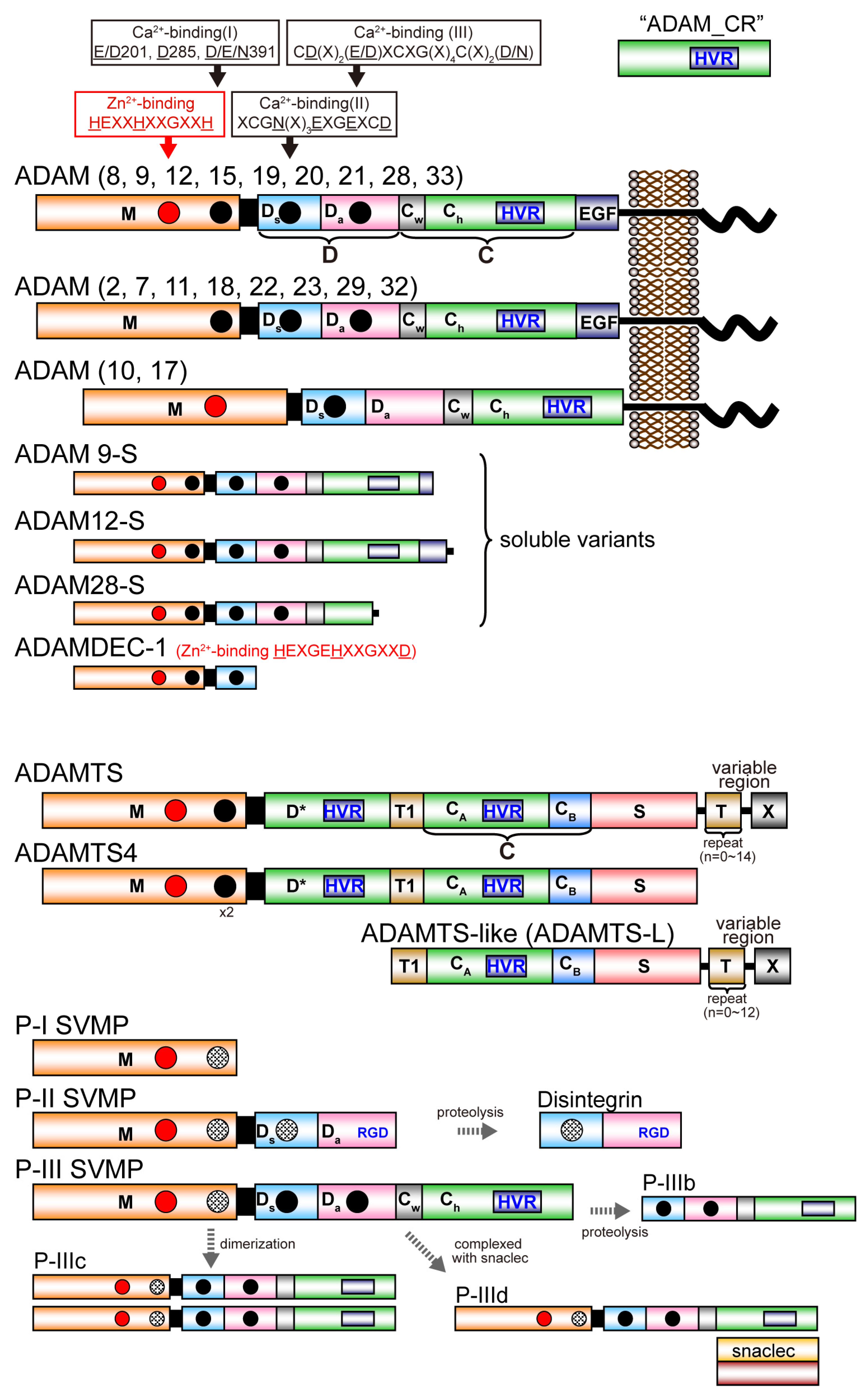
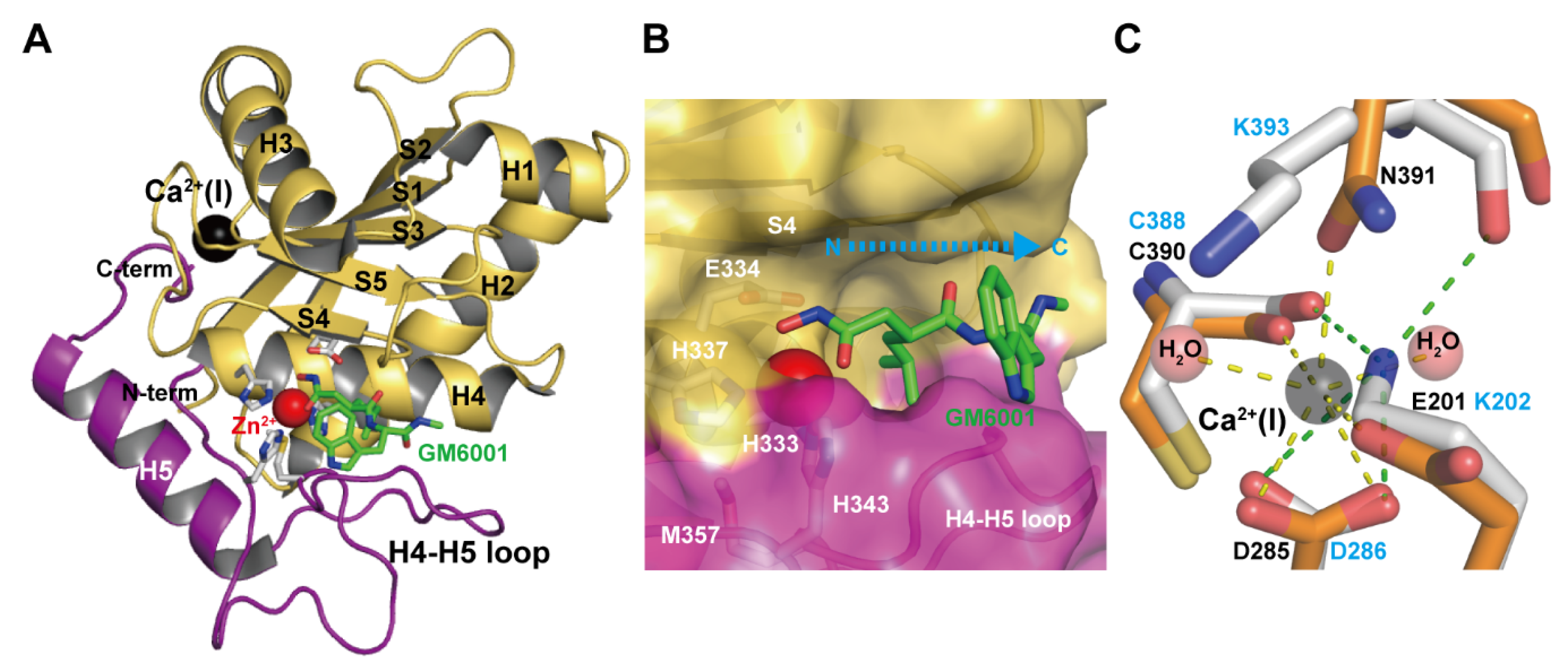
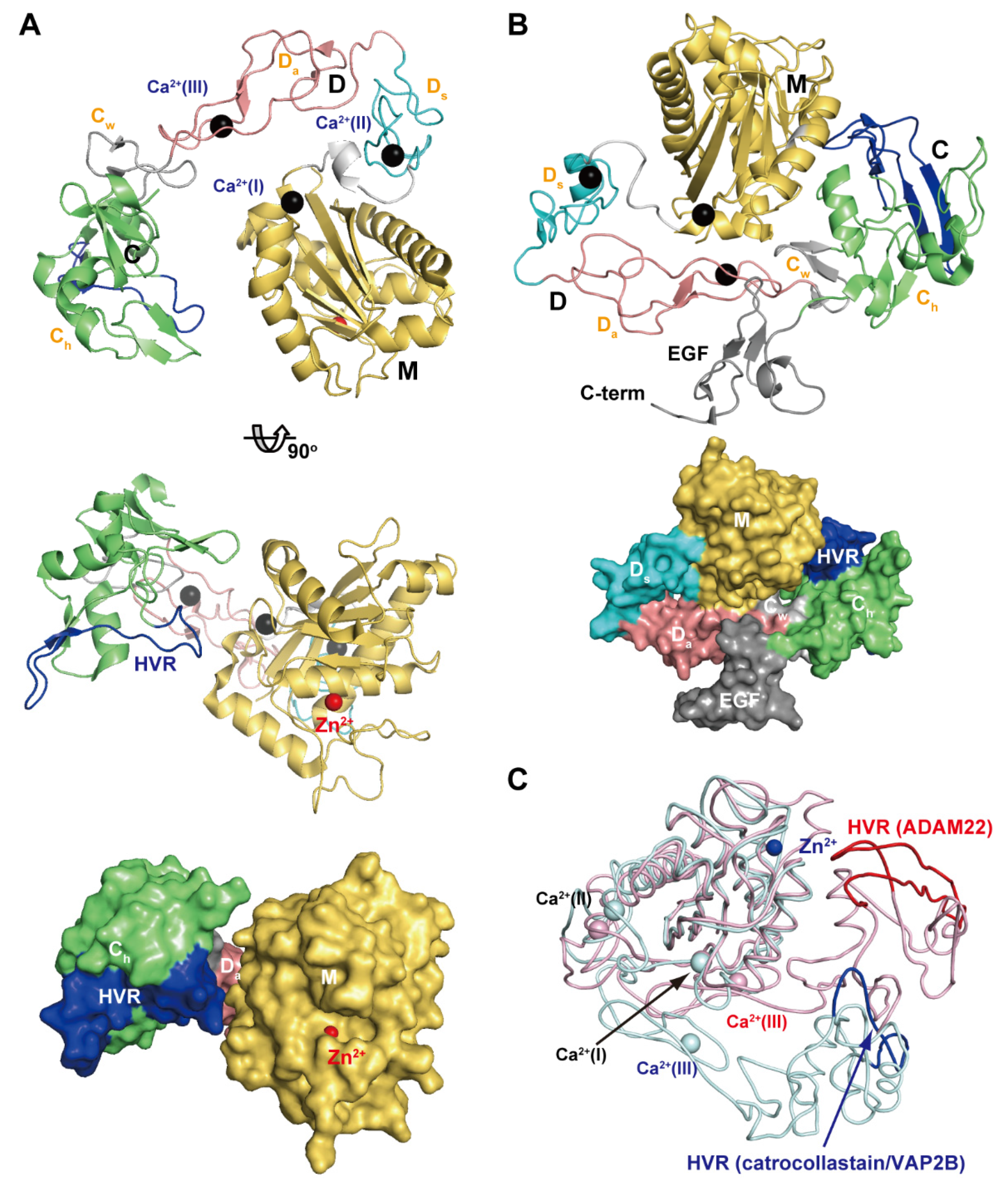
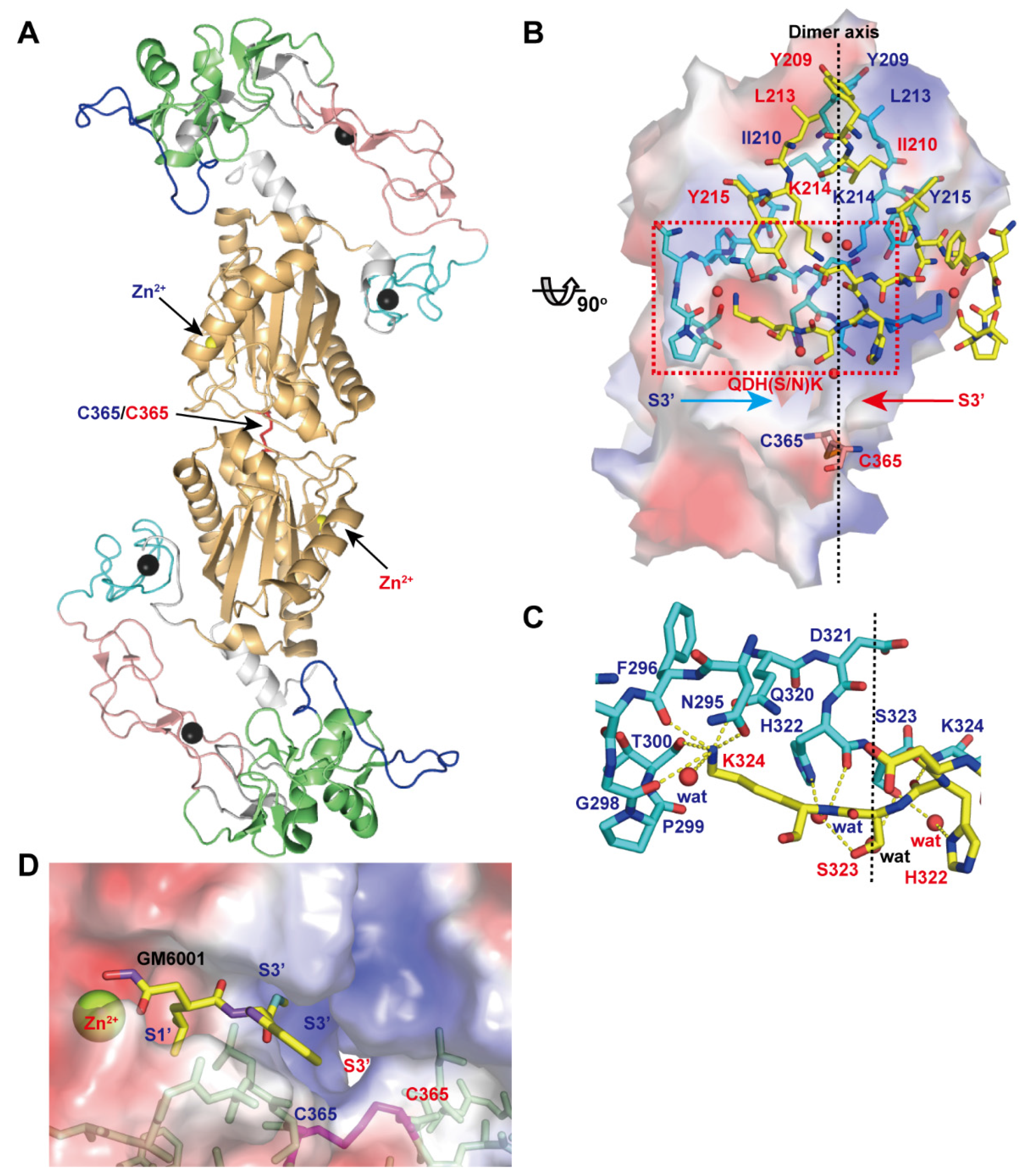
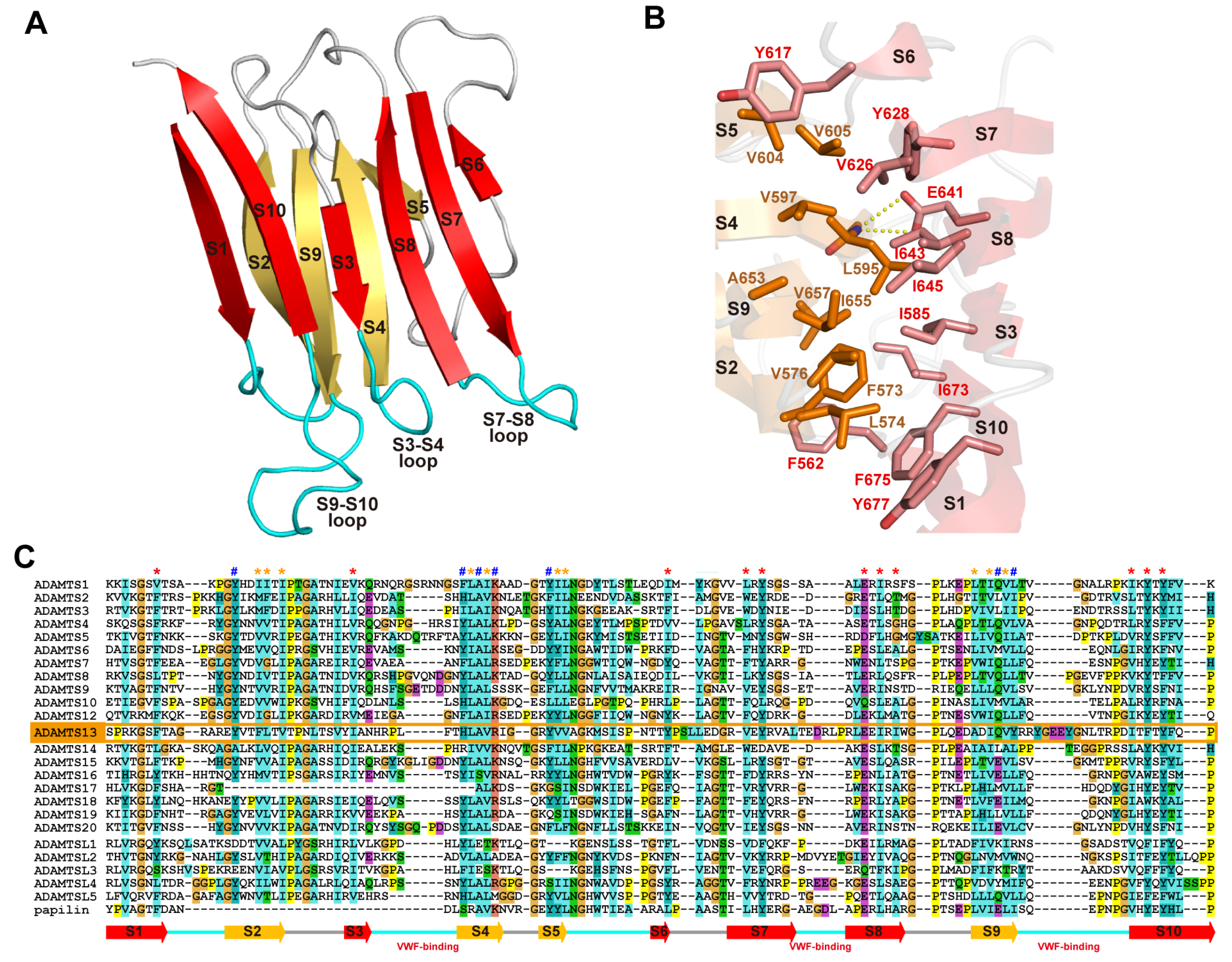
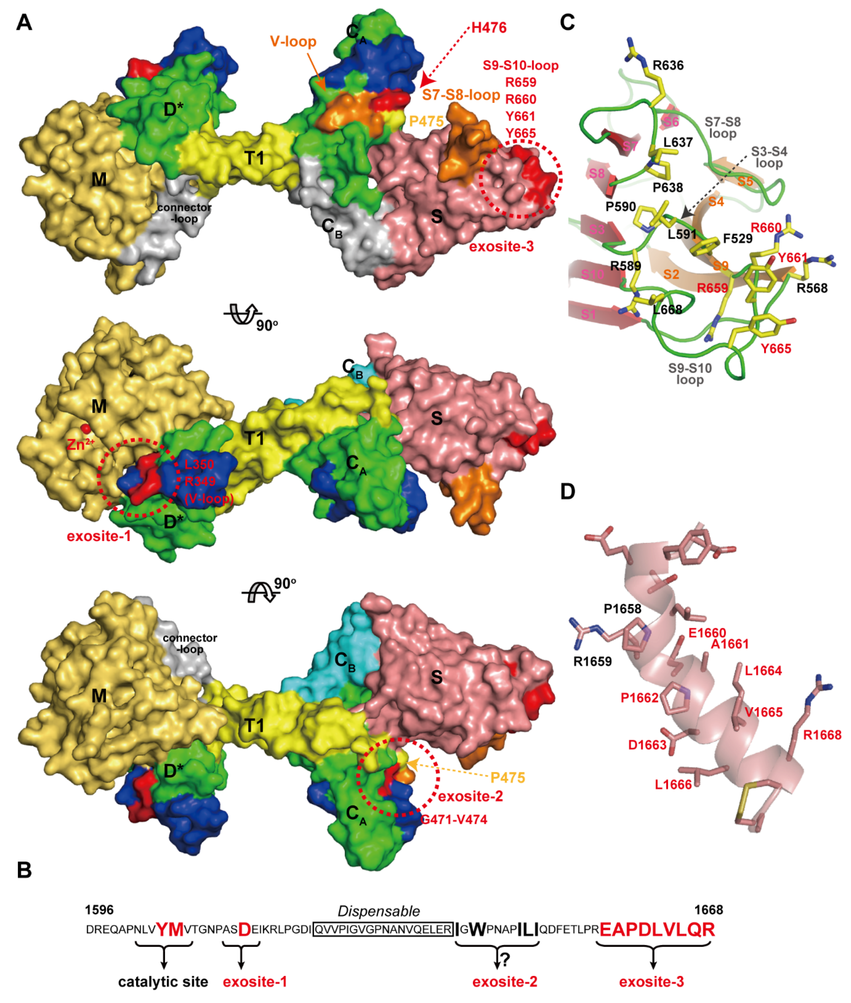
A disintegrin and metalloproteinase (ADAM) family
proteins constitute a major class of membrane-anchored multidomain
proteinases that are responsible for the shedding of cell-surface
protein ectodomains, including the latent forms of growth factors,
cytokines, receptors and other molecules. Snake venom metalloproteinases
(SVMPs) are major components in most viper venoms. SVMPs are primarily
responsible for hemorrhagic activity and may also interfere with the
hemostatic system in envenomed animals.
SVMPs are phylogenetically most
closely related to ADAMs and,
together with ADAMs and related ADAM with
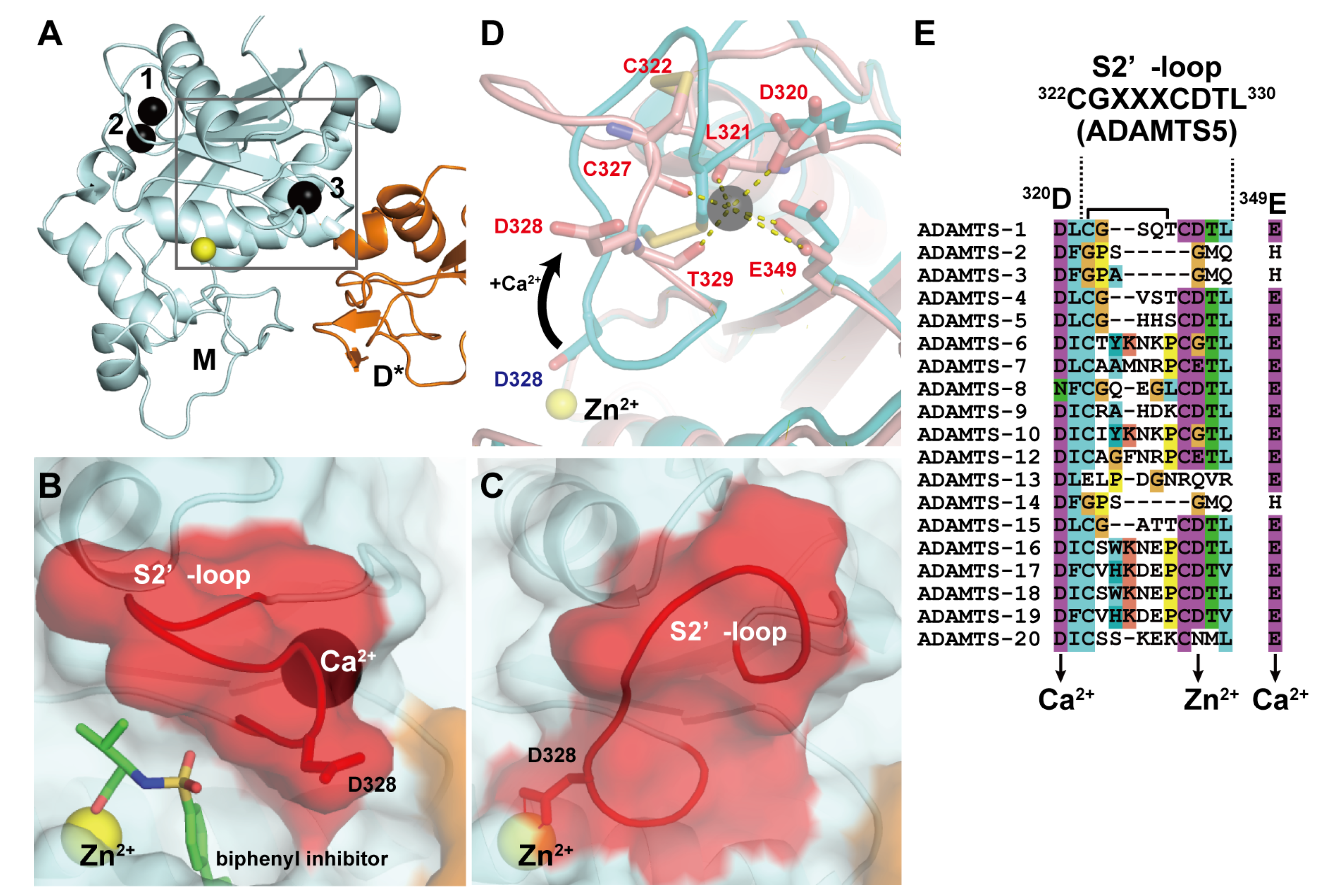
thrombospondin motifs (ADAMTS) family proteinases,
constitute
adamalysins/reprolysins or the M12B clan (MEROPS database) of
metalloproteinases.
Although the catalytic domain structure is
topologically similar to that of other metalloproteinases such as matrix
metalloproteinases, the M12B proteinases have a modular structure with
multiple non-catalytic ancillary domains that are not found in other
proteinases. Notably, crystallographic studies revealed that, in
addition to the conserved metalloproteinase domain, M12B members share a
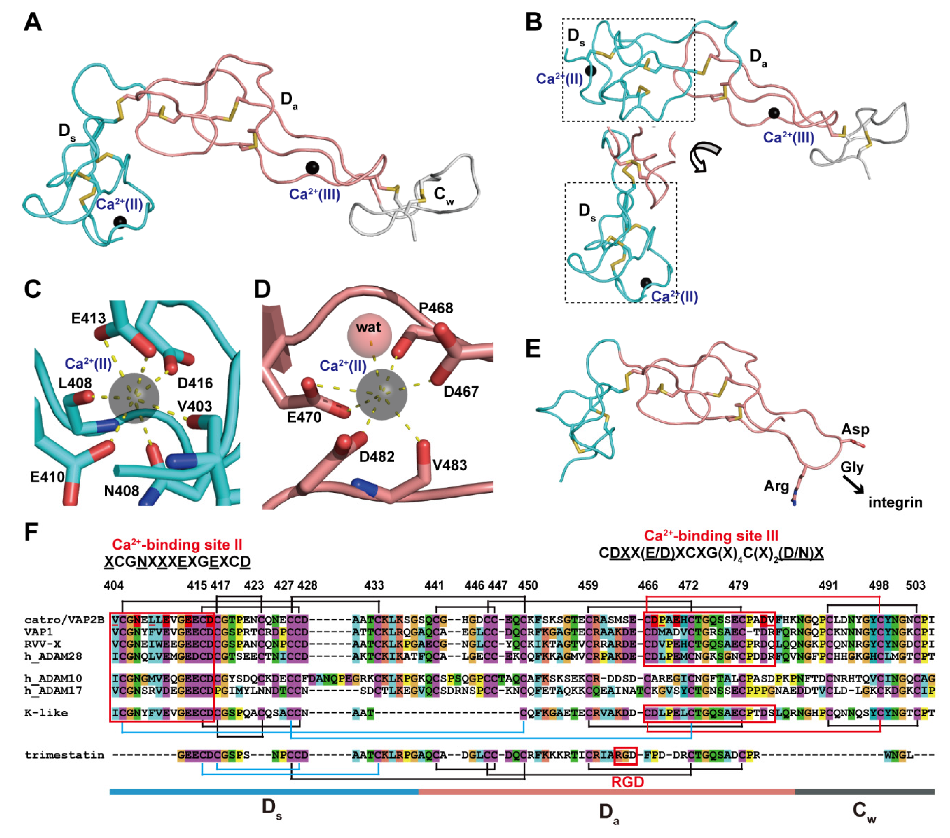
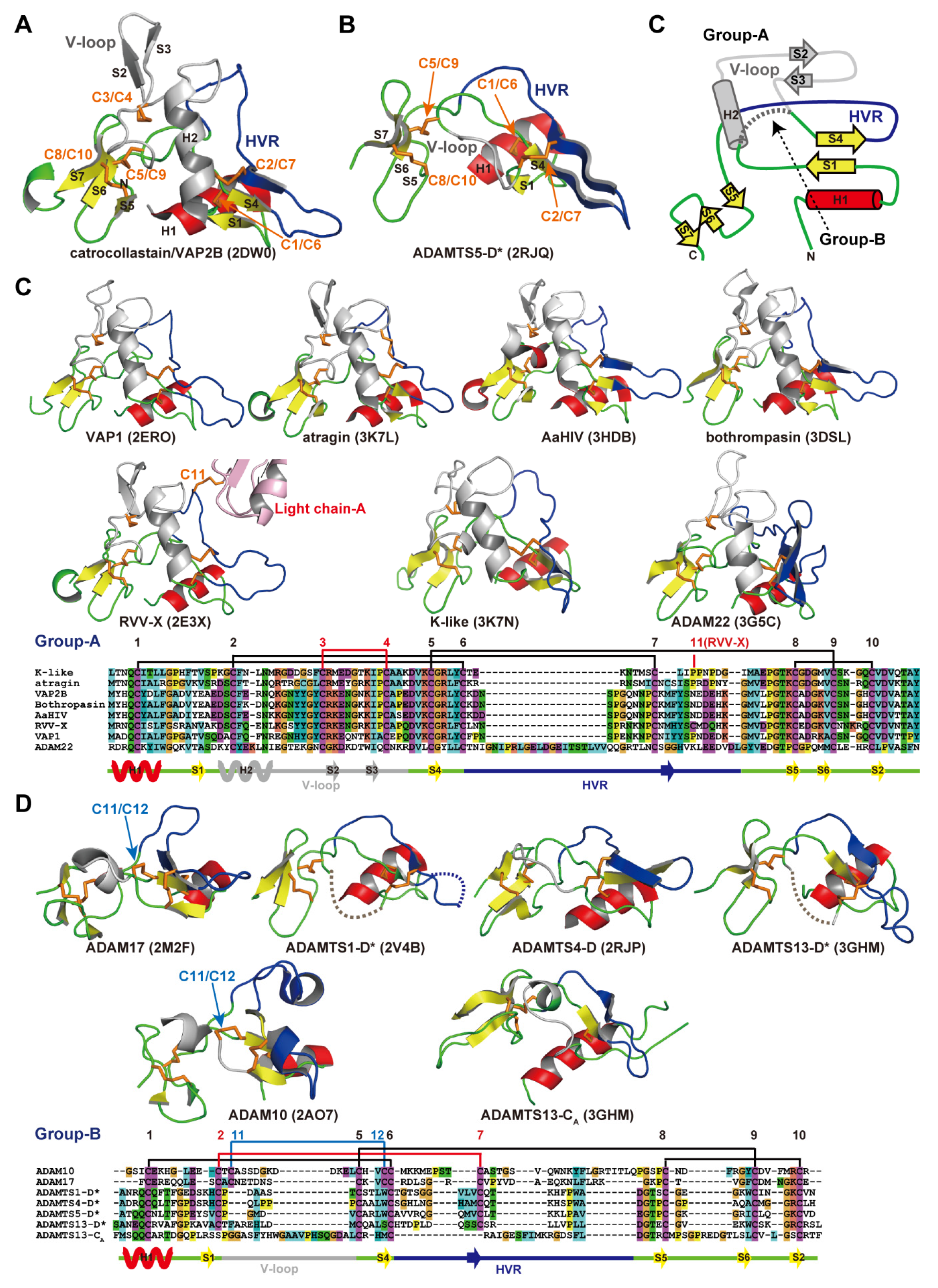
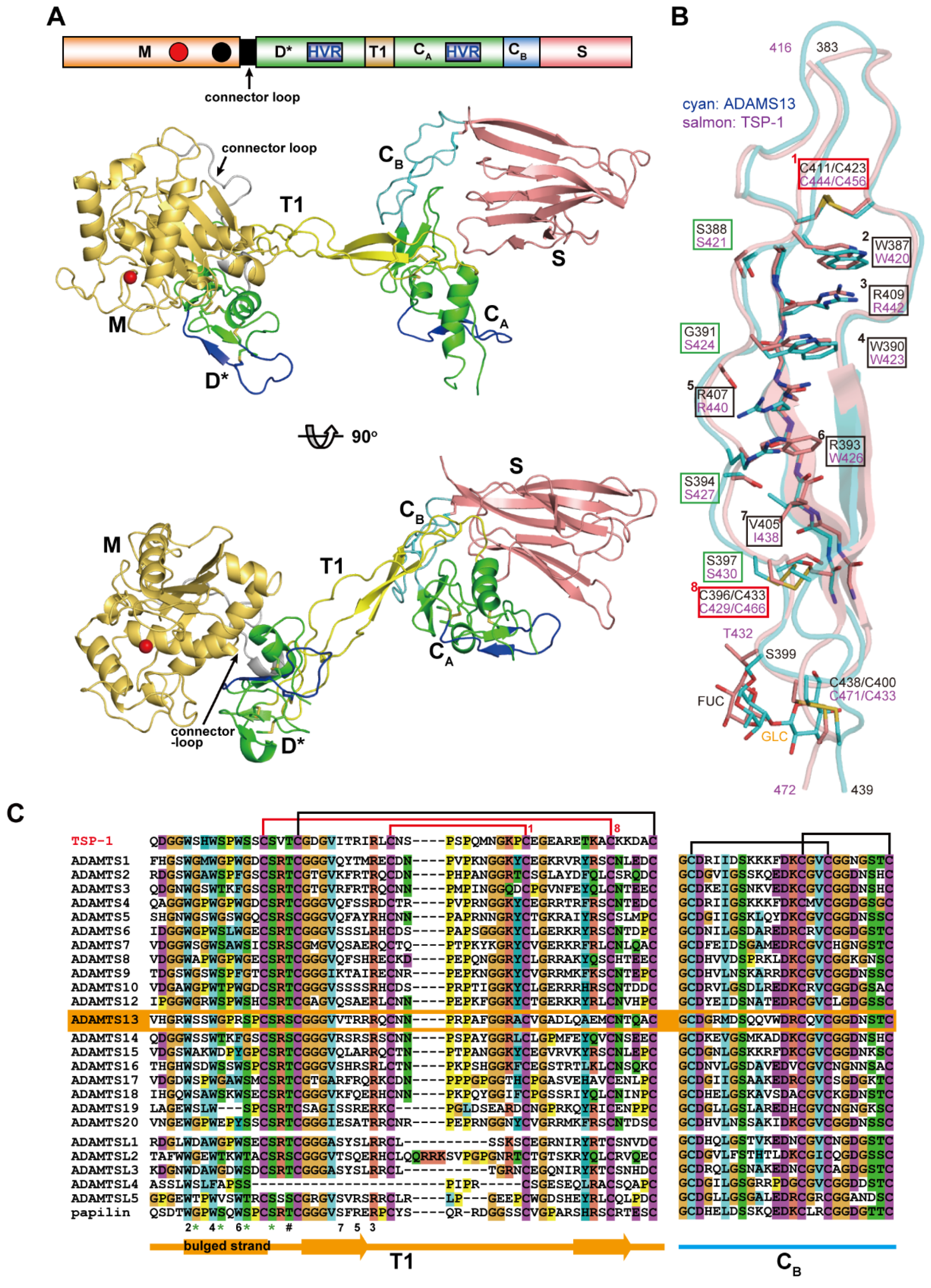
hallmark cysteine-rich domain designated as the “ADAM_CR” domain.
Despite their name, ADAMTSs lack disintegrin-like structures and instead
comprise two ADAM_CR domains.
This review highlights the current state
of our knowledge on the three-dimensional structures of M12B
proteinases, focusing on their unique domains that may collaboratively
participate in directing these proteinases to specific substrates.

